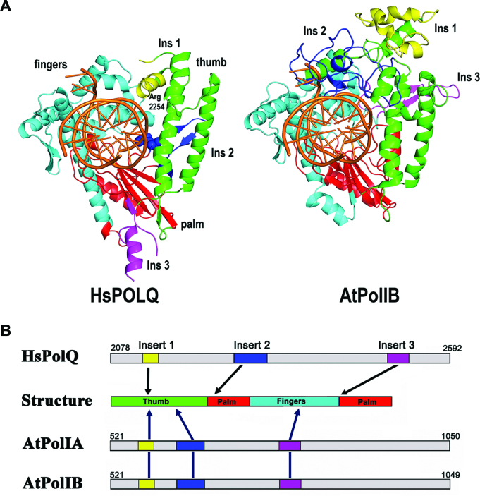
Figure 8.
AtPolIB and POLQ present structurally equivalent insertions. Structural comparison between POLQ and AtPolIB. (A) Crystal structure of POLQ showing the interaction between R2254 and the 3′ phosphate of the primer strand. No continuous electron density was observed for regions corresponding to insertions 1 and 3. dsDNA is colored in orange and the palm, thumb and fingers subdomains are colored in red, green and cyan respectively. Structural model of AtPolIB showing the structural localization of its three insertions. Insertion 3 of AtPolIB is in a similar region that insertion 2 of POLQ. (B) Graphical representation of the insertions in POLQ and AtPolIB. In Both enzymes, insertion 1 is located at the thumb subdomain. POLQ insertions 2 and 3 are located at the palm subdomain, whereas in AtPolIB they are located at the thumb and fingers subdomain respectively.

