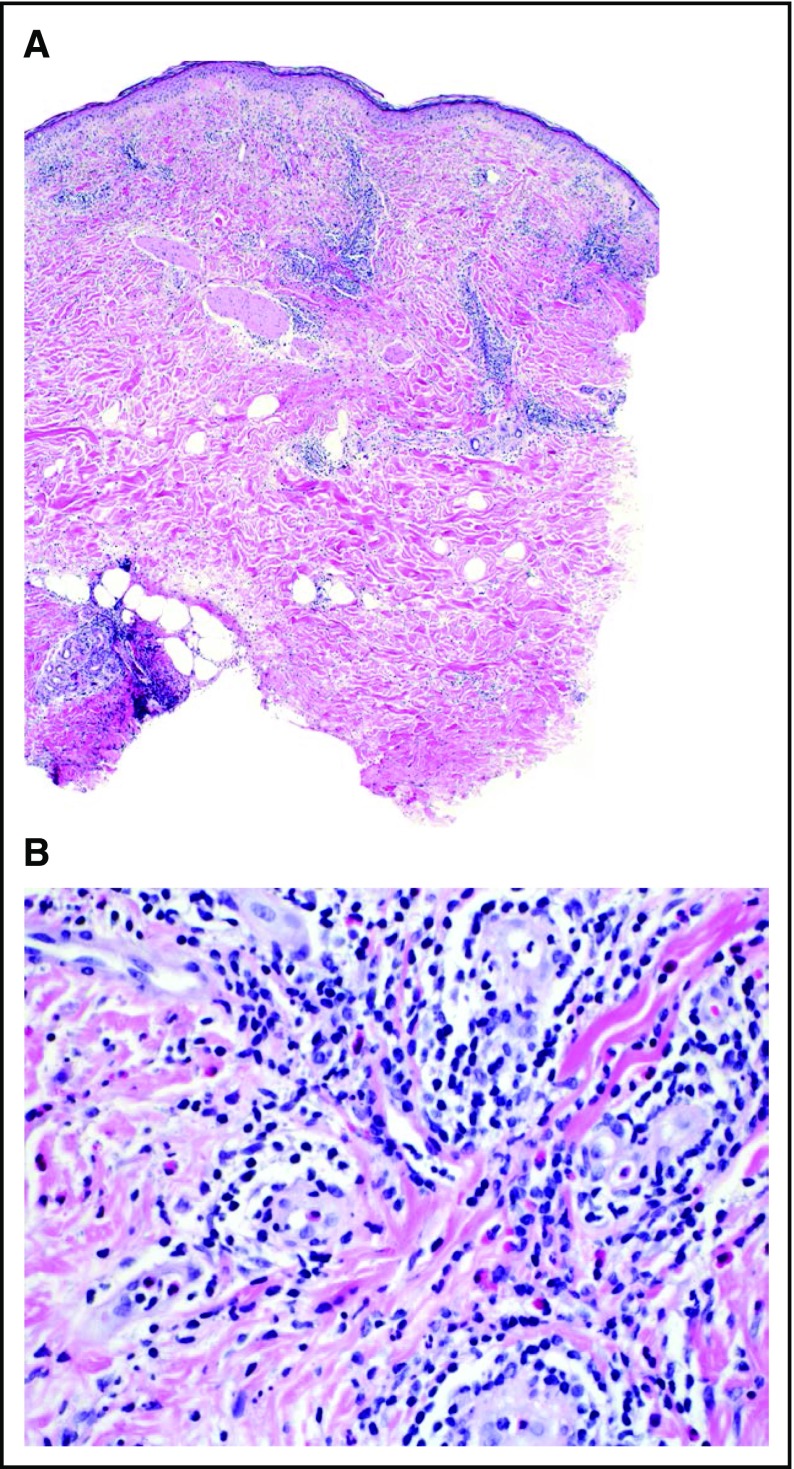
Figure 2.
Representative histopathologic response at vaccination site. (A) Hematoxylin and eosin (H&E) 4×: a fully developed reaction to vaccination. Note the prominent infiltration of lymphocytes at all levels of the skin, including the subcutaneous fat. (B) H&E 40×: higher magnification reveals numerous perivascular lymphocytes and eosinophils with perineural infiltration. The endothelial cells of the venules are swollen. There are scattered mononuclear histiocytes present in the infiltrate.

