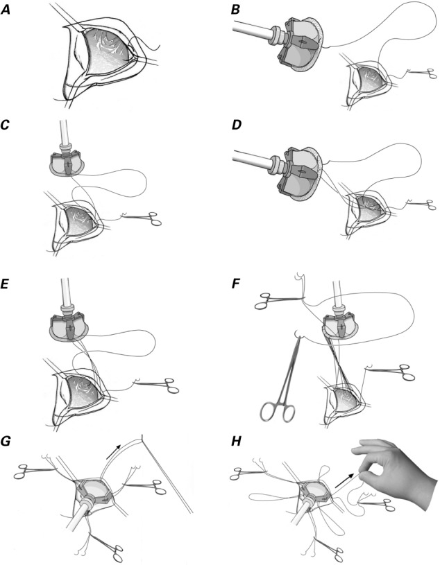
Fig. 1.

Drawings illustrate the operative technique. A) The first stitch passes through the leaflet attachment at the right coronary and noncoronary leaflet commissure. B) The 2nd stitch passes through the sewing cuff of the prosthesis. C) The 3rd stitch passes under the previous suture and is placed at the leaflet attachment. D) The 4th stitch passes over the previous suture and is placed on the sewing cuff. E) The 5th stitch passes under the previous suture and is placed at the annulus. F) The end of the first suture and one end of the 2nd suture are held with rubber-tipped forceps, which are then placed on the left shoulder of the patient. The left coronary leaflet is sutured in the same manner, with use of the other end of the suture. G) The suture at each nadir is pulled with a nerve hook. H) Six ends of 3 sutures are pulled individually, and the loops at the nadir are tightened.
