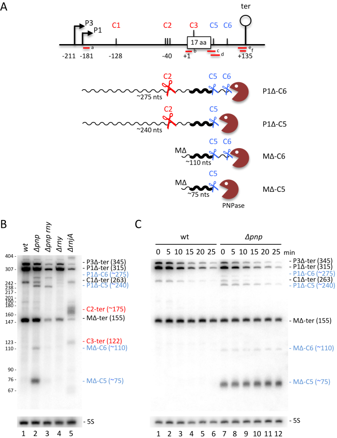
Figure 8.
Degradation of the hbsΔ mRNA can also be initiated within the 3′-UTR. (A) Schematic of the B. subtilis hbsΔ constructs showing positions of different oligonucleotide probes. Labeling is as in Figure 1. Transcripts are shown as wavy lines. Key cleavage events C2, C5, C6 are symbolized by scissors; PNPase by a brown Pacman symbol. (B) Northern blot of total RNA isolated from wt, Δpnp, Δpnp rny, ΔrnjA and Δrny strains carrying plasmids expressing the wt hbsΔ construct (n = 1). (C) Northern blot of total RNA isolated from wt and Δpnp strains carrying plasmids expressing the wt hbsΔ construct at different times after rifampicin addition (n = 1). Blots in panels B and C were probed with oligo CC1997 (hbs probe c) and then HP246 (5S rRNA) as a loading control. Degradation intermediates seen in the Δpnp strain are labeled in blue; those accumulating in the ΔrnjA strain are labeled in red.

