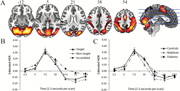
Fig. 1.
A (top): dominant 20% of loadings for Component 1 (red/yellow = positive loadings, threshold = 0.22, max = 0.32). Images are displayed in neurological orientation (left is left) with MNI z-axis coordinates. B (bottom left): mean FIR-based predictor weights plotted over post-stimulus time (discrimination conditions averaged). C (bottom right): mean FIR-based predictor weights plotted over post-stimulus time by group (task conditions averaged). FIR, finite impulse response; HDR, hemodynamic response.

