Figure 2.
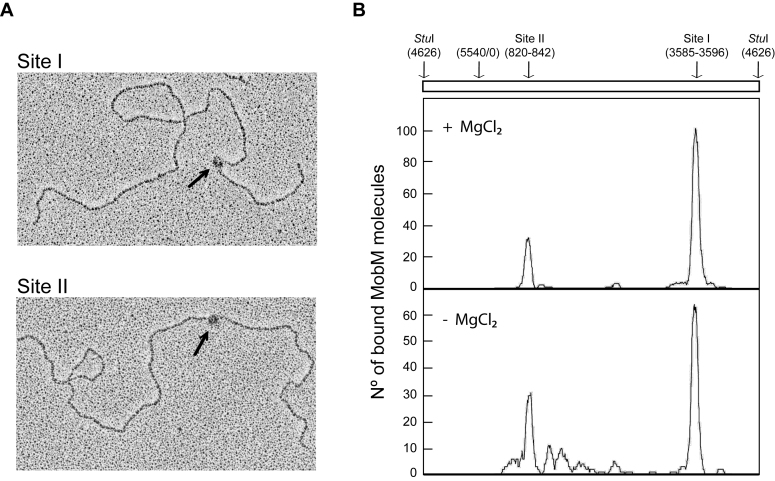
Binding of MobM to supercoiled pMV158 DNA. Plasmid pMV158 was incubated with MobM at 30°C for 30 min. MobM–pMV158 complexes were fixed with glutaraldehyde and then pMV158 was linearized with StuI. (A) Electron micrographs of linear plasmid molecules with MobM bound to site I or site II (black arrows). (B) Distribution of MobM positions on the pMV158 DNA when the binding reactions were performed in the presence of MgCl2 or in its absence. The coordinates of the two main MobM-binding sites are indicated.

