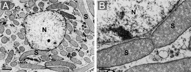
Fig. 2.
—Transmission electron microscopy (TEM) micrographs of the Sodalis endosymbiont of Henestaris halophilus. (A) Overwiew of a bacteriocyte completely filled by rod-shaped endosymbiont (S). (B) Enlarged image of dividing endosymbionts showing the characteristic gammaproteobacterial cell structure. Nucleus (N).

