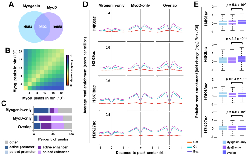
Figure 5.
Overlapping of myogenin with MyoD in histone acetylation. (A) Union analysis of MyoD and myogenin ChIP-seq signals in myoblasts differentiated for 24 h. (B) The top 20,000 MyoD and myogenin peaks were ranked by read enrichment, and grouped into bins with increments of 2000. Each box represents the fraction of sites bound by MyoD and myogenin in that bin. (C) The association of unique and overlapping MyoD and myogenin peaks with distinct chromatin states. (D) The average ChIP-seq signal profiles for H4K8ac, H3K9ac, H3K18ac and H3K27ac at poised enhancers bound by MyoD- or myogenin-only, or both in proliferating myoblasts (GM), and myoblasts differentiated for 24 h in the absence or presence bexarotene (Ctl or Bex 50 nM). (E) Quantification of log2-fold change in histone acetylation.

