Abstract
Rapidly decreasing cost of next-generation sequencing has led to the recent availability of large-scale RNA-seq data, that empowers the analysis of gene expression variability, in addition to gene expression means. In this paper, we present the MDSeq, based on the coefficient of dispersion, to provide robust and computationally efficient analysis of both gene expression means and variability on RNA-seq counts. The MDSeq utilizes a novel reparametrization of the negative binomial to provide flexible generalized linear models (GLMs) on both the mean and dispersion. We address challenges of analyzing large-scale RNA-seq data via several new developments to provide a comprehensive toolset that models technical excess zeros, identifies outliers efficiently, and evaluates differential expressions at biologically interesting levels. We evaluated performances of the MDSeq using simulated data when the ground truths are known. Results suggest that the MDSeq often outperforms current methods for the analysis of gene expression mean and variability. Moreover, the MDSeq is applied in two real RNA-seq studies, in which we identified functionally relevant genes and gene pathways. Specifically, the analysis of gene expression variability with the MDSeq on the GTEx human brain tissue data has identified pathways associated with common neurodegenerative disorders when gene expression means were conserved.
INTRODUCTION
The analysis of gene expressions via hybridization-based microarray technologies has enjoyed much success in the last two decades. Genes regulating a myriad of human diseases have been identified in microarray studies, including those for brain tumors (1), breast cancer (2–4), skin tumors (5), and a number of neurological disorders (6–8). Nonetheless, microarray experiments can be limited by the presence of cross-hybridization artifacts (9), intensity variability at low expression levels (10), signal saturation of highly expressed genes (11), and partial assessment of genes restricted to annotated transcripts (12). Utilizing next-generation sequencing (NGS) technologies, RNA sequencing (RNA-seq) has largely improved upon the limitations of microarray technologies and rapidly emerged as the preferred tool for transcriptome analysis (12). Ever decreasing costs of high-throughput sequencing have led to the recent availability of large-scale RNA-seq studies by providing datasets with moderate to large sample sizes. These include the Encyclopedia of DNA Elements (ENCODE) (13), the Genotype-Tissue Expression (GTEx) Project (14), the Genetic European Variation in Health and Disease (GEUVADIS) dataset (15), etc. The analysis of large-scale RNA-seq count data presents both new challenges and opportunities.
Gene expression variability can provide important insights on how genes function in biological processes beyond those acquired from standard analysis of gene expression means. For instance, variability analysis of gene expression levels has identified transcriptional regulators in the development of early human embryos (16). Gene expression variability at aberrant levels can suggest disruptions or dysregulations of biological processes (17,18). Recent studies have associated increased levels of expression variability with Schizophrenia (19) and aggressive chronic lymphocytic leukemia (20). A number of methods for the analysis of gene expression variability has been proposed for applications in microarray studies (21–27). However, as early RNA-seq studies often have only a limited number of samples that cannot be reliably applied to assess statistical variability, the analysis of gene expression variability has, so far, been largely ignored in RNA-seq studies that focused on the analysis of gene expression means (28–35). The availability of large-scale RNA-seq studies presents an unprecedented opportunity to evaluate gene expression variability without the encumbrance of the many limitations of microarray technologies.
The analysis of large-scale RNA-seq count data brings about several new challenges that are vital towards the analysis and interpretation of both gene expression mean and variability. Excess zeros, beyond those realized from biological variations, are often present in a significant proportion of genes in large-scale RNA-seq studies. This is often attributed to technical variations from read failures in low-count samples (36) and has been suggested to contribute to elevated levels of overdispersion in RNA-seq data (37). Furthermore, by evaluating a relatively large number of observations, outlying samples are more likely to be encountered in large-scale studies. As gene expression analysis often seeks to interrogate biologically consistent effects across treatments, it is important to identify and remove outlying observations to achieve robust biological interpretation and reproducibility of results (32,38). Most particularly, procedures for differential gene expression analysis often focus on evaluating the null hypothesis that log fold-changes (FCs) between cases and controls are exactly zero, such that there is no differentiation between cases and controls. However, given moderate to large sample sizes, the null hypothesis is often easily rejected, and this can educe a deluge of statistically significant genes, most of which are differentiated at only very modest levels that are biologically uninteresting (39,40). Effective methods are much needed to overcome these challenges in order to provide robust and biologically meaningful analysis of both gene expression mean and variability in large-scale RNA-seq studies.
In microarray studies, log-transformed intensity levels are often assumed to follow a normal or Gaussian distribution (41), and the analysis of gene expression variability usually involves evaluating the normal variances directly (24–26) or conducting heterogeneity tests under assumptions of continuous and symmetric distributions (21–23,27). Yet, RNA-seq counts are both discrete and asymmetrically distributed, such that the analysis of RNA-seq count data requires a much different approach and interpretation from those of microarray data analysis (42).
The Poisson distribution has been proposed for RNA-seq counts that approximates the binomial probability of independently sampled reads (43,44). Given an expected value of E(Y) = μ, a Poisson random variable Y manifests an intrinsic variance of Var(Y) = μ. The Poisson has been largely accepted as a suitable model for the analysis of technical replicates (45,46). However, in large-scale RNA-seq studies, investigators are typically interested in examining biological replicates at different treatment levels in order to interrogate consistent effects of treatments on gene expressions.
Individual subjects in biological replicates engender additional variability over technical ones. The coefficient of dispersion or variance-to-mean ratio ϕ = Var(Y)/μ is a standard measure of additional variability due to biological variations (26,47,48). It has been found to be advantageous in interpreting variability, free from potential finite-number effects due to varying abundances (49,50). Let Yig be the read count at subject i and gene g. We employ the mean-dispersion model in this paper based on the coefficient of dispersion, where E(Yig) = μig and Var(Yig) = ϕigμig. The variance of Yig consists of a technical μig and biological ϕig component, where μig represents the intrinsic variability due to independent sampling of reads and is biologically uninteresting whereas ϕig characterizes the additional variability arising from biological variations. Thus, interpreting the dispersion ϕig will be the focus of gene expression variability analysis in large-scale RNA-seq studies. Current procedures for RNA-seq counts often assume the negative binomial with the mean-variance relationship Var(Yig) = μig(1 + αgμig), where αg is invariant across subjects at each gene (28–30,51). Compared to the negative binomial, the mean-dispersion framework can allow for more direct interpretation of variability due to biological variations, such that a log FC in ϕig can be explicitly attributed towards a log FC in total variance where log [Var(Yig)] = log (ϕig) + log (μig). An additional advantage is that biological variabilities ϕig are allowed to vary over both individual genes and subjects, with which the power of large-scale RNA-seq studies can be exploited to incorporate dynamic and complex biological relationships. We will provide a generalized linear model (GLM) framework that can incorporate the effects of both treatments and additional covariates on the mean μig and dispersion ϕig. This allows the proposed model to account for a wide array of studies, for example, when cases and controls exhibit different variabilities and when variabilities of gene counts may be influenced by additional covariates, such as age, gender, or different stages in a biological process. In the analysis of RNA-seq data, the average expression strength at a gene has been observed to influence expression variability, where genes with decreased average counts tend to have increased overall variability (51). The mean-dispersion GLM accounts for potential changes in the baseline variability due to differences in average expression counts at each gene by incorporating a gene-wise intercept term in the GLM on dispersion. The incorporation of a dynamic variance model also allows for robust analysis of gene expression means, in addition to enabling the analysis of gene expression variability. In this paper, we present the MDSeq, an efficient toolset based on the mean-dispersion GLM for the analysis of large-scale RNA-seq studies.
The MDSeq utilizes a novel reparametrization of the negative binomial to allow for robust statistical inference and efficient computations of the mean-dispersion model. It includes several important features to address the needs of large-scale RNA-seq studies. (1) As excess zeros can distort model estimation, it is important to account for technical zero counts in order to achieve robust and biologically interpretable results. The MDSeq includes a zero-inflated GLM model, that demarcates an excess zero state for technical zeros due to sequencing failures (36) and a random state that originates all of the nonzero counts and some biological zeros due to probabilistic realizations of the random mean-dispersion model. We will demonstrate that the incorporation of excess zeros in modeling RNA-seq counts can significantly improve power in differential analysis of both gene expression mean and variability. (2) Investigators are often interested in evaluating a given set of parameters of interest. For example, it is often of interest to perform hypothesis tests on treatment effects but not necessarily on those of additional covariates, even though they may be incorporated in the GLM. Cook's distance has been proposed for outlier detection in RNA-seq data analysis (51,52). However, it measures the influences of all parameters simultaneously, such that an observation may be identified indistinguishably as an outlier regardless of whether it is influential on treatment effects or merely the additional covariates. A novel procedure is provided with the MDSeq that allows computationally efficient detection of outliers that are influential for statistical inference on user-specified sets of parameters of interest, that can comprise any or all of the treatment effects and coefficients on additional covariates. (3) It is often of interest to determine genes with differential FCs beyond a given threshold in order to facilitate post-experimental verification and provide reproducible results at measurable expression levels. A common procedure is to select differentially expressed genes that satisfy both a log FC threshold and a p-value significance level (39,40). However, the procedure is relatively ad hoc and cannot be used to determine whether differentially expressed genes satisfy the log FC threshold with statistical significance. In the MDSeq, we develop statistically rigorous procedures for hypothesis tests of both differential mean and variability of expressions at beyond given threshold levels. The development is quite different from those previously proposed for differential analysis of mean expressions (51,71). We will show that the proposed procedure is powerful while controlling type I errors.
The MDSeq is compared with a myriad of existing tools using extensive simulations for both differential expression mean and variability analyses. Results suggest that our procedures are robust and powerful in a wide spectrum of data scenarios. The MDSeq is further demonstrated on two large-scale datasets from the GTEx project, where we uncovered functionally relevant genes and gene pathways in the human skin and cerebral cortex. Excess zeros, outliers, and the need for hypothesis tests at beyond given threshold levels are illustrated on these two real datasets with the MDSeq in Table 1, Supplementary Table S2, and Table 2, respectively. We implemented the MDSeq in a user-friendly R package, freely available at https://github.com/zjdaye/MDSeq. The software allows parallel processing with multiple threads for efficient computations.
Table 1. Genes exhibiting excess zeros in the GTEx tissue data.
| With excess zeros | Without excess zeros | |
|---|---|---|
| Brain tissue | 5555 (21.4%) | 20396 (78.6%) |
| Skin tissue | 3739 (14.3%) | 22347 (85.7%) |
Numbers and percentages, in parentheses, of genes are shown. Expressions of genes are considered to have excess zeros if significance test for presence of excess zeros has p-value <0.05.
Table 2. Genes significant for differential mean and dispersion under various hypothesis tests.
| Mean only | Variability only | Both mean and dispersion | Total | |
|---|---|---|---|---|
| Cortex versus cerebellum brain tissues | ||||
| |log2FC| ≠ 0 | 7711 (29.96%) | 377 (1.46%) | 11,968 (46.51%) | 20,056 (77.94%) |
| |log2FC| > 1 | 3945 (15.33%) | 385 (1.50%) | 3214 (12.49%) | 7544 (29.32%) |
| |log2FC| > 2 | 1489 (5.79%) | 117 (0.45%) | 774 (3.01%) | 2380 (9.25%) |
| Sun-exposed versus sun-protected skin tissues | ||||
| |log2FC| ≠ 0 | 4740 (18.29%) | 1896 (7.32%) | 2757 (10.64%) | 9393 (36.25%) |
| |log2FC| > 1 | 12 (0.05%) | 53 (0.20%) | 49 (0.19%) | 114(0.44%) |
| |log2FC| > 2 | 4 (0.02%) | 7 (0.03%) | 8 (0.03%) | 19 (0.07%) |
The numbers of genes that are significant decrease with more stringent thresholds. Genes are considered significant at FDR q-value <0.05 and insignificant at FDR q-value ≥0.2. Percentages of significant genes are computed out of 25,909 and 25,734 total genes for analysis of the skin and brain tissue data, respectively.
MATERIALS AND METHODS
Mean-dispersion model
Let Yig be the read count for sample i and gene g. The MDSeq utilizes a novel reparameterization of the real-valued negative binomial Yig ∼ NB(μig, ϕig) in order to allow gene expression variability to be modeled explicitly based on the coefficient of dispersion. For notational simplicity, we assume a given gene g throughout the ‘Materials and Methods’ section and do not specify its index g in the read count Yig.
Consider the real-valued negative binomial or Pólya distribution,
 |
(1) |
where E(Yi) = μi and Var(Yi) = ϕiμi for ϕi > 1. The mean-dispersion formulation has the probability model,
 |
(2) |
where θi = θ(μi, ϕi) = μi/(ϕi − 1).
GLM has been applied to extend the classical linear model for RNA-seq counts at the mean expression level (30,34,51,53–55). In this paper, we define the mean-dispersion GLM based on log-linear relationships on both the mean and dispersion, such that
 |
(3) |
where xij and uik are design matrix elements and βj and γk are coefficients for the mean and dispersion, respectively. Intercept terms are included on both the mean and dispersion, such that the dispersion intercept allows the MDSeq to account for potential differences in baseline variability due to variations in average expression strengths at each gene (51). The design matrices {xij} and {uik} include contrasts to indicate treatments on subjects. The MDSeq allows for a number of contrast coding schemes described in Supplementary Methods. For example, in a simple case-control study, one can set xi1 = ui1 = 1 for all subjects, xi2 = ui2 = 0 for cases, and xi2 = ui2 = 1 for controls. Additional covariates describing clinical, demographic, and other experimental factors may also be included in the design matrices {xij} and {uik}. In many instances, the design matrices {xij} and {uik} can be the same. However, different contrasts and covariates can be applied in the MDSeq on the mean and dispersion, respectively, to allow applications in a wide array of studies. For example, data sources can be directly incorporated as additional factors in the dispersion GLM, if it is believed that different labs may contribute data at different quality levels and variations.
We used natural logarithms in Equation (3) according to conventions in the GLM literature (56). The natural logarithm and log2 are related through the identity log2(·) = log (·)/log (2), and, for convenience, options to output results in the log2 scale are provided in the MDSeq software. The mean-dispersion GLM can be efficiently estimated via constrained optimization techniques (57,58). Further details are provided in Supplementary Methods.
Modeling excess zero counts
Technical excess zeros are often present in a significant proportion of genes in large-scale RNA-seq data. It is often necessary to incorporate them in order to obtain interpretable results for both gene expression mean and variability analyses. We employ the zero-inflated model (59–62),
 |
(4) |
where 0 ≤ s < 1 is the probability of technical excess zeros. Equation (4) describes two states from which RNA-seq counts may arise. An excess zero state is observed with probability s that generates only zero counts, and a negative binomial state is observed with probability 1 − s that generates all of the nonzero counts and a few of the zero counts. That is, the model aims to partition zero counts probabilistically into those arising from technical variations at the excess zero state and those from biological variations at the negative binomial state.
Maximum likelihood (ML) estimates for the zero-inflated mean-dispersion GLM are computed by developing an expectation-maximization (EM) algorithm under constrained optimization (63). Detailed descriptions of algorithm are provided in Supplementary Methods.
Test to determine presence of excess zeros
We evaluate the presence of excess zeros by testing the hypothesis  against
  in the zero-inflated model of Equation (4). Under the null hypothesis when s = 0, all of the zero counts are assumed to arise from biological variations at the negative binomial state. We apply a likelihood ratio test with the statistic
  , where
  and
and  are the log-likelihoods at ML estimates of β = (β0, β1, …, βp)T and γ = (γ0, γ1, …, γq)T under hypotheses
  and
and  , respectively. The log-likelihoods
  and
and  are presented in Equations (S2) and (S4), respectively, of Supplementary Methods. Test statistic Ds follows a
  distribution under
  , from which we obtain the p-value at the tail distribution. The likelihood ratio test is applied to provide more robust inference, whereas the Wald test has been found to be sometimes unstable for inference on s in zero-inflated models (59,60). When
  is rejected, the zero-inflated model of Equation (4) is applied instead of the usual mean-dispersion GLM (Equation (1–3)) in further analyses.
Wald tests for GLM coefficients
Consider the GLM of Equation (3). We apply Wald tests to evaluate significances of the coefficients βj and γk on log mean and dispersion, respectively, with the statistics  and
and  , where
  and
and  are obtained as inverses of observed Fisher informations (64).
  and
and  follow the
  distribution under the null hypotheses βj = 0 and γk = 0, respectively, from which we obtain the p-values at the tail distributions. Wald tests for GLM coefficients can be used, for example, in evaluating significances of additional covariates on RNA-seq counts.
The observed Fisher informations for the mean-dispersion and zero-inflated mean-dispersion models are provided in Supplementary Methods, using closed-form Hessian matrices that allow for efficient computations of test statistics.
Standard tests of differential expression mean and dispersion
Denote  as the contrast vector at factor level ℓ. Then, based on Equation (S1) of Supplementary Methods, log FCs of expressions from factor level ℓ0 to ℓ1 can be estimated as
  and
and  for differential mean and dispersion, respectively.
Standard procedures consider hypothesis tests of the alternatives  for differential expression mean and
  for differential dispersion. Wald statistics Wμ for testing
  and Wϕ for testing
  are provided in Equation (S9) of Supplementary Methods, with which the p-values are obtained at
  tail distributions.
To correct for multiple hypothesis testing across genes, we apply the Benjamini–Yekutieli false discovery rate (FDR) that allows for arbitrary dependence in this paper (65).
Hypothesis tests at beyond a given log fold-change threshold
In large-scale RNA-seq studies with moderate to large numbers of samples, standard tests that evaluate the compliant hypothesis that any change in differential expressions may occur, such as by testing the alternative  , would often result in the selection of a large proportion of genes that are only mildly differentially expressed. To allow for experimental replication and interpretation of results, it is often of interest to identify genes with differential changes beyond a given threshold level. In this paper, we develop rigorous procedures based on one-sided hypothesis tests within restricted parameter spaces (66–68) and union-intersection principle (69,70). The development is quite different from those previously proposed for differential analysis of mean expressions (51,71).
We are interested in evaluating whether the differential mean or dispersion of expressions are significant beyond a given log FC threshold; in other words, we wish to test the alternative hypothesis  or
or  , respectively, for some threshold τ > 0. This is accomplished through a two-step procedure. Consider the analysis of differential expression mean. (1) Test of the alternative
  is first evaluated asunder as one-sided hypothesis tests of the alternatives
  and
and  . Wald statistics are derived under restricted parameter spaces (66–68), whereas p-values are computed for each test using mixture distributions (72–74). (2) The p-value for testing the composite alternative hypothesis
  is obtained as the minimum of p-values for testing the alternatives
  and
and  by the union-intersection principle (69,70). That is,
  is accepted if either of the alternative
  or
or  is accepted.
Details of thresholded hypothesis tests for differential mean and dispersion are provided in Supplementary Methods. We note that an asymptotically equivalent approach can also be developed based on the likelihood ratio statistics with log likelihoods maximized under restricted parameter spaces (68,74,75). However, this approach requires recomputing GLM estimates under restricted parameter spaces and is computationally more intensive.
Detection of outliers influential for inference on a given set of parameters of interest
RNA-seq data analyses often focus on a set of parameters of interest. For example, in differential expression analysis, one is mainly interested in hypothesis tests involving treatment effects of cases and controls. On the other hand, it is not necessarily of interest to interpret hypothesis tests of additional covariates, although they are often incorporated in the GLM to mitigate conditional effects. In this case, it can be advantageous to focus on the set of parameters of interest, instead of all the parameters as in the Cook's distance (51,52), for efficient detection of outliers.
Suppose that we are interested in evaluating hypotheses based on subsets  and
and  of the GLM parameters β = (β0, β1, …, βp)T and γ = (γ0, γ1, …, γq)T, respectively. For instance, in evaluating treatment factors of L levels, the parameters
  and
and  are defined as coefficients (β1, β2, …, βL − 1)T and (γ1, γ2, …, γL − 1)T, respectively, on the contrast matrix. A standard likelihood ratio test can be applied for
  and
and  with the statistic
  , where

 is the maximum log-likelihood with the coefficients of
  and
and  set equal to 0. For outlier detection, we do not consider zero-valued counts, as excess zeros are already accounted for by the zero-inflated GLM (Equation (4)).
Traditional procedures for outlier detection are often based on the leave-one-out approach (52,76). Consider the change in the likelihood ratio statistic when sample i is removed, defined as  where
where  is constructed by removing yi from
  and
and  is the likelihood ratio statistic computed without sample i. The difference of likelihood ratio statistics Ii provides a natural measure of the influence of the ith sample on inferences based on
  and
and  , such that an extreme value of Ii may suggest that sample i is an outlier. To identify all the outliers, the influence measure Ii needs to be estimated at each sample i. However, this involves computing the log-likelihoods


 and
and  repetitively with each sample i removed, which can be computationally prohibitive in large-scale RNA-seq studies.
In this paper, we propose to apply a one-step estimator for Ii based on parameter estimates computed only once on all samples (52,56,77,78). The one-step estimator  is obtained as a weighted sum of standardized deviance and Pearson residuals. Details are provided in Supplementary Methods. We compare
  to a variance-gamma distribution (79,80) and remove sample i from ensuing analyses if
  is below the (αout/2)th-quantile or above the (1 − αout/2)th-quantile of the variance-gamma, where we use αout = 0.05 in this paper. We note that the estimator
  is not guaranteed to follow a variance-gamma distribution due to potential estimation error from outliers and dependence. However, we found that the procedure works well in practice, as extreme outliers can be easily identified with the computationally efficient one-step estimator (see Results). The proposed procedure can be applied on any set of parameters of interest, including all parameters.
Data normalization and preprocessing
Normalization is necessary to account for technical biases of read counts at different samples, such as those due to varying sequencing depths. In this paper, we applied the trimmed mean of M values (TMM) procedure, that has been found to be robust against technical biases (30,81). We adjusted the raw counts by using TMM normalization factors provided by the TMM procedure together with the library sizes. Adjusted counts were subtracted by 0.5 and then raised to the smallest integers for the downstream expression analysis. The MDSeq also allows the user to apply other normalization factors, including the relative log expression (RLE) (29), upper-quartile (46), and conditional quantile normalization (cqn) (82) factors. In addition to normalized counts, the MDSeq also provides an option to offset sample-specific normalization factors in the GLM with raw counts. Further details are provided in Supplementary Methods.
RESULTS
MDSeq performs the best for gene expression variability analysis in large-scale studies
We compared the MDSeq with six other methods that have been proposed for variance heterogeneity analysis in microarray gene expression studies. Bartlett's test is a classical procedure for evaluating unequal variances between groups (22,23,83). The Levene's test is a robust alternative to Bartlett's under non-normal data (22,23,27,84–87). It is further improved upon for data with outlying samples by using the trimmed-mean, in which we removed the top 10% outlying samples (88). The heteroscedastic regression, that extends the simple linear regression with nonconstant error variances, has been proposed for detecting genetic loci controlling gene expression variability in microarray studies (24–26,87). We apply the heteroscedastic regression to include additional covariates in this paper; specifically, a normal probability model  is applied where μi and
  are linear and log-linear in treatment factors and additional covariates, respectively. The mean-absolute-deviation (MAD) test has been proposed as a robust procedure for differential variability analysis of microarray gene expressions (21,89). Moreover, the Fligner–Killeen test utilizes a nonparametric approach for variance comparisons (90). Using the R programming language, we applied the Bartlett's test with the bartlett.test function, Levene's tests with leveneTest from the car package, heteroscedastic regression with dglm using the Gaussian family and log-link options, our implementation of the MAD according to Ho et al. 2008 (21), and the Fligner–Killeen test with fligner.test.
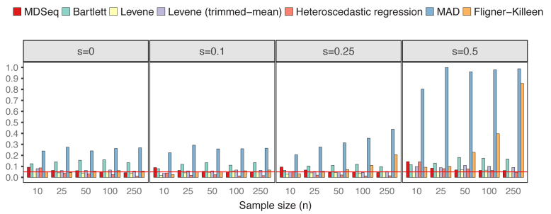
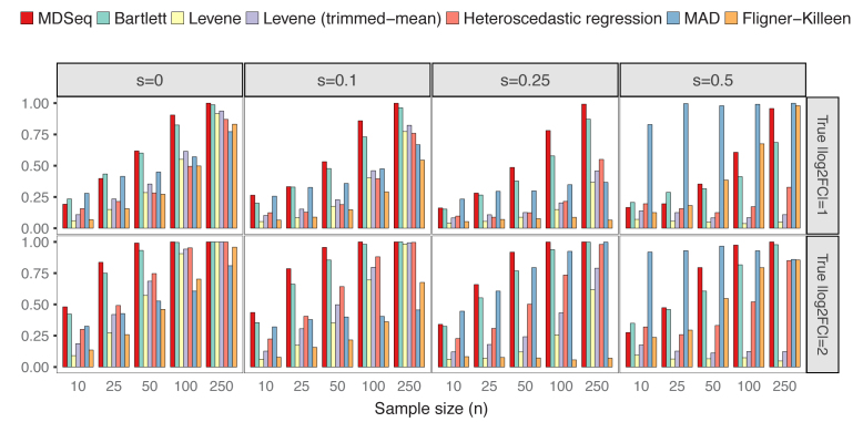
Differential variability analysis is often applied in order to obtain additional insights beyond those already acquired from standard differential analysis of gene expression means. Thus, in this study, we focused on evaluating scenarios when gene expression means are consistent across cases and controls. To compare with Bartlett's, Levene's, MAD, and Fligner–Killeen tests that do not allow the incorporation of additional covariates, we simulated count data from NB(μ0, ϕ0) for controls and NB(μ0, 2log2FCϕ0) for cases without additional covariates, where we set μ0 = exp (5) and ϕ0 = exp (4) as the constant mean and baseline dispersion, respectively. Excess zeros were incorporated according to the zero-inflated model (see ‘Materials and Methods’ section). Both empirical powers and type I errors for each procedure were estimated as proportions of p-values < 0.05 from 1,000 repetitions, where type I errors were computed under constant variances over cases and controls with log2FC = 0. Figure 1 presents type I errors at varying probability of excess zeros and sample sizes. The MDSeq, Levene's tests, and heteroscedastic regression have well controlled type I errors at around the theoretical level of 0.05, except when sample sizes are extremely small. On the other hand, both Bartlett's and MAD tests have inflated type I errors in all scenarios with the worst performance at s = 0.5 when the probability of excess zeros is relatively large. This suggests that the Bartlett's test, that tends to be sensitive to departure from normality (84), and the MAD test may be inappropriate for RNA-seq count data, especially when excess zeros are present. The Fligner-Killeen test has well controlled type I errors at s small but can have inflated type I errors at s large, suggesting that excess zeros can effect the nonparametric approach. Figure 2 shows that the MDSeq dominates Levene's tests and heteroscedastic regression in terms of power, when |log2FC| = 1 and |log2FC| = 2, in all scenarios, whereas the MDSeq dominates the Fligner-Killeen test except at s large when the Fligner-Killeen test can have inflated type I errors. Further, performances of Levene's tests and heteroscedastic regression, that do not incorporate excess zeros, quickly deteriorate with increasing probability of excess zeros s, whereas the MDSeq remains robust at s large. These results suggest that the MDSeq, based on a zero-inflated GLM count model, can be advantageous for variability analysis of large-scale RNA-seq count data. Scenarios with additional covariates are presented in Supplementary Figures S1 and S2 for type I errors and powers, respectively.
Figure 1.
Type I errors in the absence of differential expression variability. There are n samples of cases and controls each and varying proportions of excess zeros s. The MDSeq, Levene's tests, and heteroscedastic regression have well controlled type I errors for moderate to large sample sizes, whereas Bartlett's and MAD tests have highly inflated type I errors. Results are based on 1,000 simulations without additional covariates. Reference lines (in red) are drawn at the 0.05 error rate.
Figure 2.
Powers of detecting differential expression variability. There are n samples of cases and controls each and varying proportions of excess zeros s and log2 fold-changes log2FC. The MDSeq often performs the best when sample sizes are moderate or large. Levene's tests and heteroscedastic regression tend to deteriorate in performance with increasing proportions of excess zeros s. Results are based on 1,000 simulations without additional covariates.
MDSeq is advantageous for mean expression analysis of counts with excess zeros
The MDSeq was compared with six other methods for differential mean analysis. The DESeq2 (29,51) and edgeR (30,34,53–55) are popular procedures for RNA-seq analysis at the mean level. Similar to the MDSeq, these methods were developed based on negative-binomial regressions. We compared with the edgeR using both conditional maximum likelihood (ML) (55) and quasi-likelihood (QL) (31,91) estimates with the robust option (34). The voom from the limma package (33,35) circumvents modeling of count data directly by applying the linear model on log-transformed RNA-seq counts. The tweeDEseq (92,93) applies a general family of Poisson-Tweedie probability models to better account for heavy tails and scenarios when the amount of excess zeros is modest, whereas these properties of RNA-seq counts were modeled directly using a zero-inflated dispersion model in the MDSeq. The ShrinkBayes (37,94) incorporates zero-inflated negative binomial models via a Bayesian framework.
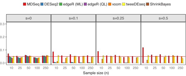
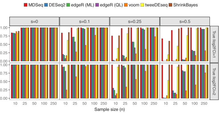
We simulated count data without additional covariates from NB(μ0, ϕ0) for controls and NB(2log2FCμ0, ϕ0) for cases, where μ0 = exp (5) and ϕ0 = exp (4). Excess zeros were incorporated according to the zero-inflated model (see ‘Materials and Methods’ section). Powers and type I errors were estimated as proportions of p-values < 0.05 from 1,000 repetitions, except that Bayesian false discovery rates (computed with the BFDR function in ShrinkBayes by setting the multcomp option to FALSE) were used for results with the ShrinkBayes (37,94). Figure 3 presents type I errors based on scenarios when gene expression means are consistent across cases and controls. The MDSeq, voom and tweeDEseq generally control type I errors well at around 0.05, while, at extremely small sample sizes, the MDSeq has moderately inflated type I errors. The DESeq2 and edgeR methods control type I errors well at around 0.05 when no excess zeros are present (s = 0), whereas type I errors are nearly 0 when s > 0, suggesting that they may be conservative under the presence of excess zeros. Powers are presented in Figure 4. All methods are comparable in terms of power at s = 0 when no excess zeros are present. However, the performances of DESeq2, edgeR methods, and voom quickly deteriorate under the presence of excess zeros s > 0. This suggests that unaccounted technical zeros may thwart the detection of differential expression means with these methods. The tweeDEseq, that applies a general probability model, performs relatively well under the presence of excess zeros. Nonetheless, the MDSeq and ShrinkBayes, that directly model technical zeros, dominate the tweeDEseq in terms of power when the proportion of excess zeros s is large. This suggests the need to account for technical excess zeros directly using zero-inflated GLMs.
Figure 3.
Type I errors in the absence of differential expression means. There are n samples of cases and controls each and varying proportions of excess zeros s. The MDSeq controls type I errors well at moderate to large sample sizes. DESeq2 and edgeR methods may be conservative under the presence of excess zeros s > 0. Results are based on 1,000 simulations without additional covariates. Reference lines (in red) are drawn at the 0.05 error rate.
Figure 4.
Powers of detecting differential expression means. There are n samples of cases and controls each and varying proportions of excess zeros s and log2 fold-change log2FC. The MDSeq and ShrinkBayes often perform the best among methods compared under the presence of excess zeros s > 0. Results are based on 1,000 simulations without additional covariates.
Supplementary Table S1 provides computational times for all methods. The MDSeq can compute for 1,000 simulations in about half a minute or less in all cases. It is faster than tweeDEseq and ShrinkBayes. Although employing a more involved EM algorithm, the MDSeq is faster than DESeq2 at large sample sizes, whereas it is slower than edgeR and voom.
Supplementary Figures S3 and S4 present type I errors and powers, respectively, under scenarios when additional covariates are present. Moreover, Supplementary Figures S5 and S6 present type I errors and powers, respectively, when both expression means and variability are nonconstant across cases and controls. In this scenario, voom has highly inflated type I errors at s = 0 when no excess zeros are present. Further, type I errors increase with increasing sample sizes. This suggests that the voom, although it performs well under equal variances between cases and controls, may be inadequate for RNA-seq count data arising from heterogeneous count distributions. This may be due to the voom relying on a symmetric, normal distributional model (33,35,95) on log-transformed counts, which can be asymmetrically distributed (42). We note that a weighted regression approach may be helpful for mean expression analysis under heteroscedasticity with voomWithQualityWeights (96).
MDSeq provides valid hypothesis tests to evaluate absolute log fold changes above a given threshold
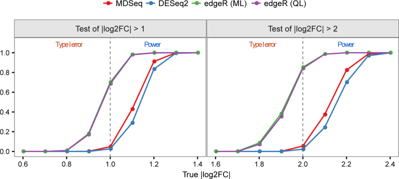
We compared hypothesis tests to evaluate absolute log FCs above given thresholds of expression means for the MDSeq, DESeq2 with the lfcThreshold option, and edgeR using the glmTreat function. The DESeq2 and edgeR do not incorporate technical zeros and were not developed for gene variability analysis. Thus, we did not generate excess zeros and focused on differential expression means in this comparison. In Figure 5, the left panel evaluates the hypothesis Ha: |log2FC| > 1, whereas the right panel considers Ha: |log2FC| > 2. We see that the MDSeq controls type I errors well at around or below the 0.05 theoretical level for Ha: |log2FC| > 1 (Ha: |log2FC| > 2) when the underlying log FC |log2FC| ≤ 1 (|log2FC| ≤ 2). On the other hand, edgeR has inflated type I errors when the underlying absolute log FC is at or moderately less than the given threshold. Moreover, the MDSeq dominates DESeq2 in terms of power when the underlying log FC is above the given threshold. For example, at test of |log2FC| > 2 when the true |log2FC| = 2.2, the MDSeq has a power of 0.824 while the DESeq2 has a power of 0.701, that represents an improvement of over 17.5% for the MDSeq. Nonetheless, DESeq2 can be validly applied in most scenarios, as it does not incur inflated type I errors. We note that the MDSeq can further incorporate excess zeros and evaluate hypotheses involving expression variability.
Figure 5.
Hypothesis tests to evaluate absolute log fold-changes of expression means above given thresholds. Type I errors are shown at |log2FC| less than or equal to a given threshold, and powers are presented when |log2FC| is greater than the given threshold. The MDSeq and DESeq2 have well controlled type I errors, whereas edgeR methods have highly inflated type I errors when |log2FC| is at or moderately less than the given thresholds. The MDSeq has greater power than DESeq2 when |log2FC| is moderately above the given thresholds. There are 500 samples of cases and controls each. Results are based on 1,000 simulations generated from NB(2log2FCμ0, ϕ0) with μ0 = exp (5) and ϕ0 = exp (4) for varying log2FC. No excess zeros were generated with s = 0. Reference lines (in gray) are drawn at the corresponding threshold levels.
Supplementary Figure S7 presents inequality hypothesis tests for differential variability analysis with the MDSeq. Hypothesis tests to evaluate absolute log FCs of expression variability have well controlled type I errors, while powers increase more gradually above the given threshold levels compared with those of hypothesis tests of expression means.
Computationally efficient detection of outliers influential on a set of parameters of interest
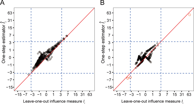
Figure 6 examines the accuracy of the one-step estimator  versus the leave-one-out influence measure Ii, obtained by repeatedly computing likelihood estimates with each sample i removed. The one-step estimator
  closely reflects the true influence measure Ii when no outliers are present (Figure 6A), whereas it is slightly less accurate under the presence of outliers (Figure 6B). We see that inaccuracy due to using a one-step estimation usually does not effect the identification of outliers, which requires influence measures to have fairly large magnitudes at the extreme tails of the variance-gamma distribution. This allows the one-step estimation procedure to provide robust yet computationally efficient outlier detection in large-scale RNA-seq studies. Supplementary Figure S8 illustrates a scenario when there are no outliers influential on treatment effects of cases and controls while observations are indiscriminately identified as outliers when influence on all parameters is considered.
Figure 6.
Accuracy of the computationally efficient one-step estimator  . The computationally efficient one-step estimator
  is compared with the leave-one-out influence measure Ii under scenarios when (A) there are no outliers and when (B) outliers are present. There are n = 250 samples each for cases and controls. Counts were generated from NB(μi, ϕi) for controls and NB(2log2FCμi, ϕi) for cases with log2FC = 2, where μi = exp (5 + (xi1 + xi2)/2) and ϕi = exp (4 + (xi1 + xi2)/2). Additional covariates xi1 and xi2 were simulated from binomial distributions Binom(2, prob = (0.5, 0.5)). In (B), five samples were randomly replaced by outliers simulated from Pois(exp (5)exp (4)). Non-outlying samples (in black) and outliers (in magenta) are plotted. Reference lines (in dashed blue) are drawn at the (αout/2)th- and (1 − αout/2)th-quantile of the variance-gamma distribution with αout = 0.05. A diagonal reference line (in solid red) is drawn at equality of
  and Ii. In (B), all five outliers were identified by the one-step estimator
  .
.
Analysis of the GTEx brain and skin tissue data
The Genotype-Tissue Expression (GTEx) project provides an expansive repository of large-scale RNA-seq data across tissue types (97). Recent studies have examined tissue-specific gene expression changes at the mean level using the GTEx data (98,99). However, studies have not been conducted, as far as we know, that effectively examined and accounted for differential changes in gene expression variability across tissue types. In this section, we illustrate applications of the MDSeq on two large-scale RNA-seq studies that compared the expression profiles of brain tissues from the cerebral cortex (obtained from 96 subjects) against those from the cerebellum (103 subjects) and profiles of skin tissues from sun-exposed (302 subjects) against those from sun-protected (196 subjects) epidermises. RNA-seq read counts were obtained from the GTEx Portal (http://www.gtexportal.org, dbGaP Accession: phs000424.v6.p1). Read counts of 26,800 and 26,144 genes with >0.05 average count per million reads across all samples were retained for gene expression analysis in the brain and skin tissue data, respectively. We note that this is a very lenient filtering criterion applied in our data analyses. In practice, a more robust criterion would require a certain number of samples to be above a given count per million reads for both the case and control groups. Further details on data preprocessing strategies are provided in Supplementary Methods. Raw counts were normalized using the trimmed mean of M values (TMM) method (81). Ensuing analyses were performed using the normalized read counts (see ‘Materials and Methods’ section.)
Statistical tests were performed on the brain and skin tissue data to determine if counts of individual genes contain excess zeros (see ‘Materials and Methods’ section). About 21% of individual genes from brain tissues and 14% of genes from skin tissues were found to exhibit significant excess zeros (Table 1). Moreover, estimated proportions of technical zeros can be relatively large for these genes (Supplementary Figure S9). Thus, we see that incorporating excess zeros can be crucial toward the analysis and interpretation of RNA-seq data. Next, outliers were identified at each gene based on the influence statistics  with respect to coefficients of contrasts (see ‘Materials and Methods’ section). About 67% of genes from brain tissues and 19% of genes from skin tissues were found to contain at least one outlier at the αout = 0.05 level (Supplementary Table S2). Samples identified as outliers were removed before further analyses were performed.
Traditional significance tests for Ha: |log2FC| ≠ 0 and composite hypothesis tests with respect to a given log FC for Ha: |log2FC| > 1 and Ha: |log2FC| > 2 were performed. Multiple hypotheses were adjusted using the conservative Benjamini–Yekutieli false discovery rate (FDR) that accounts for arbitrary dependence (65). Due to relatively large sample sizes in these studies, classical tests for no change in expression levels are easily rejected. Hypothesis tests for Ha: |log2FC| ≠ 0 on the mean and dispersion of RNA-seq counts are significant for about 78% and 36% of genes from the brain and skin tissues, respectively, whereas composite hypothesis tests with respect to a log FC Ha: |log2FC| > 1 are significant for about 29% and 0.44% of genes from the brain and skin tissues, respectively (Table 2). Volcano plots depicting p-values based on these hypothesis tests are shown in Supplementary Figures S10 and S11 for differential mean and variability, respectively, of the skin tissue data and Supplementary Figures S12 and S13 for differential mean and variability, respectively, of the brain tissue data. Moreover, a number of genes were found to be significant for age and gender covariates (Supplementary Table S3). The MDSeq, based on the GLM, allows for more interpretable results by accounting for potential biological relationships due to additional covariates. Further analyses and interpretation of results are presented as follows.
Differential variability analysis of sun-exposed and sun-protected skin tissues uncovers relevant genes overlooked by mean expression analysis
Table 3 presents genes differentially expressed in the mean or variance. Composite hypothesis tests were performed with respect to at least a two FC in either the mean or dispersion, and the Benjamini–Yekutieli false-discovery rate (FDR) was applied for multiple testing control under arbitrary dependence (65). A myriad of genes in Table 3 that are significant for differential variability but not differential expressions at the mean level are related to sun exposure of skin tissues. For example, studies have shown that ultraviolet radiations can lead to functional irregularities among genes from the histone family (HIST1H1C, HIST1H1E, HIST1H2AE, HIST1H2BG, HIST1H3D, HIST1H3H) (100,101). Keratin is an important structural material in the formation of the epidermis, and disruptions to genes of the keratin family (KRT17P1, KRT39, KRT41P, KRT6B) have been found to cause several skin disorders, including the development of carcinomas (102,103). Moreover, genes of the heat-shock protein family A (HSPA5,HSPA6) have been shown to effect cell responses to ultraviolet irradiation in human skin tissues (104–106). Absolute log FCs tend to be relatively small for these genes at mean expression levels, which may be caused by the fact that sun-exposures on subjects are not severe enough to trigger a significant change in expressions at the mean or that expression means are conserved for these functionally important genes. Results suggest that the analysis of gene expression variability can be a useful addition to traditional differential gene expression analysis at the mean level and can provide an important component towards the interrogation of gene functionality and genetic effects of human disorders. Supplementary Figures S14 and S15 provide p-values from methods at subsets of decreasing sample sizes of the genes found in Table 3, according to an approach from van Wieringen and van de Wiel (107). Full results of significance tests on all genes for both the skin and brain tissues data are provided in Supplementary Data.
Table 3. Genes significant for differential mean and dispersion of sun-exposed versus sun-protected skin tissues with respect to the threshold |log2FC| > 1.
| Gene | Ensembl Gene ID | Differential mean | Differential variability | ||||
|---|---|---|---|---|---|---|---|
| log2FC | Statistics | FDR q-value | log2FC | Statistics | FDR q-value | ||
| Genes significant for differential mean but not differential dispersion | |||||||
| C10orf99 | ENSG00000188373.4 | 1.41 | 22.61 |

|
1.49 | 7.15 | 1.00 |
| FAM83A | ENSG00000147689.12 | −1.66 | 38.80 |

|
−1.56 | 8.67 | 1.00 |
| LHFPL3-AS1 | ENSG00000226869.2 | −1.57 | 22.88 |

|
−1.65 | 9.18 | 1.00 |
| NELL2 | ENSG00000184613.6 | 1.67 | 42.67 |

|
1.30 | 2.62 | 1.00 |
| RP11-252C15.1 | ENSG00000254813.1 | 1.52 | 29.58 |

|
1.66 | 12.82 | 3.27 × 10−01 |
| RP11-371I1.2 | ENSG00000215808.2 | 1.69 | 32.25 |

|
1.20 | 0.79 | 1.00 |
| RP11-529A4.7 | ENSG00000255305.1 | 1.56 | 19.03 |

|
1.54 | 4.13 | 1.00 |
| SIX1 | ENSG00000126778.7 | 1.57 | 24.14 |

|
1.33 | 2.25 | 1.00 |
| SNORA75 | ENSG00000206885.1 | −1.43 | 21.21 |

|
−1.06 | 0.09 | 1.00 |
| STMN2 | ENSG00000104435.9 | 1.41 | 18.91 |

|
1.65 | 11.87 | 5.22 × 10−01 |
| VGLL2 | ENSG00000170162.9 | 1.70 | 26.33 |

|
1.35 | 2.58 | 1.00 |
| ZNF385B | ENSG00000144331.14 | −1.73 | 90.40 |

|
−1.42 | 5.43 | 1.00 |
| Genes significant for differential variability but not differential mean | |||||||
| AC003958.2 | ENSG00000234859.1 | −1.04 | 0.06 | 1.00 | −2.04 | 18.98 |

|
| AC018442.1 | ENSG00000235683.1 | 1.26 | 6.16 | 1.00 | 1.96 | 21.55 |

|
| ACKR2 | ENSG00000144648.10 | 1.14 | 3.46 | 1.00 | 1.86 | 22.98 |

|
| ACTC1 | ENSG00000159251.6 | 1.36 | 7.82 | 1.00 | 1.94 | 18.50 |

|
| ALOX15B | ENSG00000179593.11 | −0.54 | 0.00 | 1.00 | −1.99 | 26.32 |

|
| APOC1 | ENSG00000130208.5 | −1.01 | 0.00 | 1.00 | −1.78 | 17.61 |

|
| AWAT1 | ENSG00000204195.3 | −1.78 | 13.93 | 3.14 × 10−01 | −2.37 | 23.61 |

|
| CBLN2 | ENSG00000141668.5 | −1.76 | 13.09 | 4.75 × 10−01 | −2.30 | 22.55 |

|
| CPHL1P | ENSG00000240216.3 | −1.41 | 5.69 | 1.00 | −2.54 | 33.46 |

|
| CTB-36O1.7 | ENSG00000244921.2 | 1.24 | 2.22 | 1.00 | 2.87 | 35.56 |

|
| EEF1A1P11 | ENSG00000228502.1 | 0.11 | 0.00 | 1.00 | 1.79 | 20.11 |

|
| FADS1 | ENSG00000149485.12 | −0.91 | 0.00 | 1.00 | −2.06 | 26.78 |

|
| FAR2 | ENSG00000064763.6 | −0.66 | 0.00 | 1.00 | −1.86 | 17.94 |

|
| GPRC5D | ENSG00000111291.4 | −1.78 | 10.20 | 1.00 | −2.25 | 18.97 |

|
| HGD | ENSG00000113924.7 | −1.15 | 1.52 | 1.00 | −2.00 | 24.81 |

|
| HIST1H1C | ENSG00000187837.2 | −0.90 | 0.00 | 1.00 | −1.92 | 23.89 |

|
| HIST1H1E | ENSG00000168298.4 | −1.14 | 1.36 | 1.00 | −1.85 | 18.59 |

|
| HIST1H2AE | ENSG00000168274.3 | −1.15 | 1.84 | 1.00 | −2.50 | 58.83 |

|
| HIST1H2BG | ENSG00000187990.4 | −0.99 | 0.00 | 1.00 | −2.01 | 26.00 |

|
| HIST1H3D | ENSG00000197409.6 | −1.27 | 5.81 | 1.00 | −1.96 | 21.90 |

|
| HIST1H3H | ENSG00000203813.4 | −1.10 | 0.71 | 1.00 | −2.09 | 24.74 |

|
| HRK | ENSG00000135116.5 | 1.43 | 12.06 | 7.45 × 10−01 | 2.27 | 39.48 |

|
| hsa-mir-6723 | ENSG00000237973.1 | 0.57 | 0.00 | 1.00 | 2.09 | 32.26 |

|
| HSD17B2 | ENSG00000086696.6 | −0.99 | 0.00 | 1.00 | −1.99 | 26.22 |

|
| HSPA5 | ENSG00000044574.7 | −0.74 | 0.00 | 1.00 | −1.76 | 18.28 |

|
| HSPA6 | ENSG00000173110.6 | −0.91 | 0.00 | 1.00 | −2.26 | 42.12 |

|
| ID1 | ENSG00000125968.7 | −1.10 | 0.90 | 1.00 | −1.88 | 20.41 |

|
| KRT17P1 | ENSG00000131885.12 | −1.42 | 6.33 | 1.00 | −2.14 | 18.97 |

|
| KRT39 | ENSG00000196859.3 | −1.71 | 11.89 | 7.93 × 10−01 | −2.12 | 18.85 |

|
| KRT41P | ENSG00000225438.1 | −1.75 | 8.84 | 1.00 | −2.74 | 22.08 |

|
| KRT6B | ENSG00000185479.5 | −1.19 | 2.20 | 1.00 | −1.89 | 20.09 |

|
| MC5R | ENSG00000176136.4 | −1.51 | 7.97 | 1.00 | −2.29 | 17.52 |

|
| MIR22HG | ENSG00000186594.8 | −0.59 | 0.00 | 1.00 | −1.81 | 18.87 |

|
| MOGAT2 | ENSG00000166391.10 | −1.49 | 12.14 | 7.22 × 10−01 | −2.11 | 27.50 |

|
| MTND2P28 | ENSG00000225630.1 | 0.42 | 0.00 | 1.00 | 2.09 | 34.06 |

|
| MYH3 | ENSG00000109063.10 | 0.45 | 0.00 | 1.00 | 2.20 | 46.74 |

|
| PDE6A | ENSG00000132915.6 | −1.67 | 12.54 | 6.09 × 10−01 | −2.25 | 22.33 |

|
| PDZK1 | ENSG00000174827.9 | −0.93 | 0.00 | 1.00 | −2.00 | 26.96 |

|
| PLIN5 | ENSG00000214456.4 | −0.95 | 0.00 | 1.00 | −1.79 | 17.28 |

|
| RP11-206M11.7 | ENSG00000244468.1 | −1.04 | 0.03 | 1.00 | −2.52 | 17.45 |

|
| RP11-325K4.3 | ENSG00000261270.1 | −1.16 | 2.45 | 1.00 | −1.79 | 17.33 |

|
| RP11-325P15.2 | ENSG00000230832.3 | −1.55 | 5.79 | 1.00 | −2.99 | 23.82 |

|
| RP11-38H17.1 | ENSG00000254366.2 | 1.37 | 2.58 | 1.00 | 1.92 | 17.07 |

|
| RP11-829H16.3 | ENSG00000258525.1 | −1.46 | 12.18 | 7.22 × 10−01 | −2.28 | 40.13 |

|
| RP11-845M18.6 | ENSG00000257829.1 | −1.48 | 3.82 | 1.00 | −2.25 | 17.59 |

|
| RP11-849I19.1 | ENSG00000263146.2 | 1.04 | 0.08 | 1.00 | 1.74 | 21.42 |

|
| RP4-555D20.2 | ENSG00000261786.1 | 1.21 | 4.52 | 1.00 | 1.85 | 21.00 |

|
| RP5-857K21.7 | ENSG00000229344.1 | 0.71 | 0.00 | 1.00 | 2.66 | 57.96 |

|
| RPL29P14 | ENSG00000241112.1 | −1.43 | 12.32 | 6.77 × 10−01 | −2.13 | 28.36 |

|
| SLC6A1 | ENSG00000157103.6 | −1.29 | 12.99 | 4.96 × 10−01 | −1.78 | 18.75 |

|
| SNORD3A | ENSG00000263934.2 | −1.12 | 0.96 | 1.00 | −1.95 | 22.95 |

|
| SNORD3D | ENSG00000262202.3 | −1.43 | 7.82 | 1.00 | −2.25 | 27.69 |

|
| TRIM55 | ENSG00000147573.12 | −1.63 | 11.89 | 7.93 × 10−01 | −2.40 | 25.54 |

|
Genes are considered significant at FDR q-value <0.05 and insignificant at FDR q-value ≥0.2. Significant FDR q-values are boldfaced. The log2 fold-change log2FC, test statistics, and FDR q-value with respect to the threshold |log2FC| > 1 are shown. Positive log2FC indicates over-expression in the sun-exposed skin tissue, and negative log2FC indicates over-expression in the sun-protected skin tissue.
Gene expression variability reveals functionally important pathways in the cerebral cortex
We examine significances of gene-set pathways in terms of differential mean and variability via gene-set enrichment analysis (GSEA) (108). GSEA is performed with the software GSEA v2.2.0, obtained from http://software.broadinstitute.org/gsea, using default parameters. Genes are ranked using Wald's statistics derived from the null hypothesis H0: log2(FC) = 0 for differential mean and dispersion. This allows us to account for lowly expressed or low variance genes with potentially significant FCs in the mean or dispersion, respectively (109). GSEA using all potentially differential genes can provide an important ancillary analysis, that complements gene-by-gene significance analysis with respect to a given threshold level.
Table 4 presents enriched pathways in terms of differential mean or variability for the brain tissue data. Normalized enrichment score (NES) accounts for both differences in gene-set sizes and correlations between gene sets, whereas the FDR q-value is estimated based on the NES and is adjusted for gene-set sizes and dependency (108). Out of 1,003 pathways, two exhibited enrichment in terms of expression means only, six exhibited enrichment in terms of variability only and 90 exhibited enrichment in both mean and dispersion.
Table 4. Significantly enriched pathways for differential mean and dispersion of cortex versus cerebellum brain tissues.
| GO term | Ontology | No. genes | Differential mean | Differential variability | ||
|---|---|---|---|---|---|---|
| NES | FDR q-value | NES | FDR q-value | |||
| Pathways significant for differential mean but not differential dispersion | ||||||
| Organelle Localization | BP | 23 | 1.67 |

|
1.28 | 0.207 |
| Positive Regulation Of Phosphate Metabolic Process | BP | 21 | 1.68 |

|
1.25 | 0.243 |
| Pathways significant for differential variability but not differential mean | ||||||
| Cell Cell Adhesion | BP | 74 | 1.23 | 0.275 | 1.66 |

|
| Galactosyltransferase Activity | MF | 15 | 1.15 | 0.377 | 1.64 |

|
| Hemopoietic Or Lymphoid Organ Development | BP | 67 | 1.14 | 0.405 | 1.59 |

|
| Hemostasis | BP | 39 | 1.30 | 0.216 | 1.59 |

|
| Kinase Regulator Activity | MF | 42 | 1.29 | 0.222 | 1.62 |

|
| Protein Kinase Regulator Activity | MF | 36 | 1.30 | 0.216 | 1.65 |

|
Pathways are considered significant at FDR q-value <0.05 and insignificant at FDR q-value ≥0.2. Significant FDR q-values are boldfaced. The normalized enrichment score (NES) and FDR q-value for NES are shown. Positive NES indicates enrichment in the cortex, and negative NES indicates enrichment in the cerebellum.
Pathways enriched for gene expression differential variability often indicates functional differences between the cerebral cortex and the cerebellum. Cell-cell adhesion is foundational in the reorganization and assembly of neural circuits (110). Enrichment of cell-cell adhesion pathway may suggest increased variation and plasticity of the cerebral cortex relative to the cerebellum (111). Galactosyltransferase activity functions to catalyze the transfer of galactose to acceptor molecules. Chronic injection and build-up of D-galactose in mice have been used to model neurodegeneration and brain aging in pharmacological research (112–114). A study has noted that galactosyltransferase activity decreases progressively after birth in the cortex but not the cerebellum in mice, suggesting that galactosyltransferase activity is relatively stable in the cerebellum but variant in the cortex (115).
Moreover, pathways enriched for gene expression variability in the cortex have been found to be associated with common neurodegenerative disorders effecting the cerebral cortex, such as Alzheimer's disease and dementia. Hemopoietic or lymphoid organ development involves the progression of hematopoiesis or hemopoiesis by differentiation. It is an important process in the regeneration of brain tissues. Enrichment of hemopoietic development may indicate increased variation of tissue regenerations in the cerebral cortex relative to the cerebellum. The hematopoietic system has been associated with and proposed as target for treatment of Alzheimer's disease (116,117). Hemostasis involves the avoidance or arrest of bleeding by mediating the circulation of blood. The brain has been known to possess a sophisticated hemostasis regulatory system that protects itself from hemorrhagic injury (118). Enrichment of hemostasis variability may reflect a functional reaction to microbleeding in the cerebral cortex. Studies have found strong correlation between cerebral microbleeding and leukoaraiosis or diseases effecting cerebral white matters (116,119) and Alzheimer's disease (120). Kinase regulator activity and protein kinase regulator activity have been known to play important roles in memory and learning (121). Aberrations in the regulation of kinases have been shown to contribute towards the development of Alzheimer's disease (122–124). Therapeutic strategies targeting protein kinases of the central nervous system have been proposed (125).
On the other hand, pathways significantly enriched in terms of mean expression instead of expression variability are involved in essential biological processes that, due to evolutionary pressure, are rarely observed to contribute to common neurological disorders. Organelle localization involves essential processes of transportation and maintenance of organelles in regions of the cell that are important to normal functions of neurons. Moreover, regulation of phosphate metabolic processes is a primary means of energy regulation. For example, phosphorylation of glucose, the most significant source of energy in the brain, is needed to initiate essential pathways in the usage and storage of glucose.
Pathways significant only for differential mean or differential dispersion for the skin data are provided in Supplementary Table S4. Full GSEA results for both the skin and brain tissues data are available in Supplementary Data.
DISCUSSION
In this paper, we have presented the MDSeq that offers an efficient and comprehensive solution set for the analysis of gene expression means and variability in large-scale RNA-seq studies. The MDSeq utilizes a novel likelihood-based approach to incorporate the mean-dispersion model in a GLM framework. It introduces a zero-inflated GLM to account for technical excess zeros frequently encountered in RNA-seq data. A new approach is developed for detecting outliers influential on a user-specified set of parameters of interest, that is computationally efficient. Further, statistically rigorous hypothesis tests for gene expression differences beyond given threshold levels are provided for both differential analyses of gene expression means and variability, that allow differentially expressed genes to be identified with statistical significance at biologically interesting levels. The MDSeq has been shown with extensive simulation studies to be advantageous for the analysis of gene expression variability on large-scale RNA-seq data and for the analysis of gene expression means of RNA-seq counts with technical excess zeros. The MDSeq has been shown to perform well in the simulation scenarios considered for n ≥ 25. Applications of the MDSeq on the analyses of the GTEx skin and brain tissues data have identified functionally relevant genes and gene pathways. In particular, gene variability analysis of the human brain tissue data has revealed pathways associated with common neurodegenerative disorders, such as Alzheimer's disease and dementia.
The mean-dispersion model applied in the MDSeq is related to the quasi-Poisson, that considers the mean-variance relationship E(Y) = μ and Var(Y) = ϕμ for any positive ϕ > 0 (56,91,126,127). The quasi-Poisson is developed based on the quasi-likelihood in order to avoid the difficulty of building a probability likelihood model for count data when ϕ can assume an arbitrary positive value (91). In this paper, we focused on modeling the mean-variance relationship Var(Yig) = ϕigμig for over-dispersed data when ϕ > 1. A probability likelihood model is developed using a novel approach based on a reparametrization of the negative binomial (see ‘Materials and Methods’ section). This allows the MDSeq to take advantage of an array of theoretical results and techniques from maximum likelihood theory. For example, the one-step estimator proposed for outlier detection is based on theoretical approximations of probability likelihoods. Moreover, we note that under-dispersion is rarely encountered in RNA-seq studies and can often be attributed to extreme proportions of excess zeros (128). The mean-dispersion model naturally accounts for and qualifies our model for over-dispersion ϕig > 1 using the negative binomial, whereas technical excess zeros are evaluated and demarcated using the zero-inflated GLM. These features allow the proposed model to be robust for the analysis of RNA-seq count data.
The MDSeq variance model Var(Yig) = ϕigμig is motivated from the coefficient of dispersion ϕig = Var(Yig)/μig, that has been found to be advantageous in evaluating additional variability under varying abundances (49,50). Another measure often used in evaluating additional variability is the coefficient of variation  or coefficient of variation squared
  (129–131). The MDSeq could potentially be developed based on the mean-variance relationship
  motivated from the coefficient of variation. We note that the reparametrization of the negative binomial in the MDSeq can be analogously developed to attain a probability likelihood model for the alternative variance relationship when ηig > 1. However, investigators are usually interested in the analysis of gene expression variability in order to interrogate additional information beyond those already acquired in the standard analysis of gene expression means. Thus, differential variability analysis is often most informative when mean expression levels are consistent across treatments. In this scenario, as μig is undifferentiated, the analysis of gene expression variability would be unaffected by the choice of the mean-variance relationship.
The MDSeq utilizes a zero-inflated GLM to account for excess zeros in Equation (4). We note that the probabilistic framework is agnostic to the sources of excess zeros. It only requires estimations of the overall probability of excess zeros at a given gene in order to provide robust inference on biological variations at the random negative binomial state. For example, the MDSeq is expected to remain relatively stable when proportions of excess zeros are different across cases and controls but the overall proportions of excess zeros are the same, especially at moderate to large sample sizes (Supplementary Tables S5 and S6).
In this paper, we focused on differential analyses of gene expression means and variability due to the prevalence of case-control data in large-scale RNA-seq studies and their importance towards the identification of functional impacts of genes. The analysis of variance quantitative trait loci (QTLs) that associates genetic variants with quantitative traits has drawn much attention in recent literature (22–26,87,132–135). Variance QTLs can be a source of gene expression variability and play an important role in the genetic regulation of complex traits. The identification of variance QTLs can also help to uncover interactions among genetic variants due to the increased variability of traits influenced by genetic interactions. The MDSeq can be directly applied for the analysis of variance QTLs with RNA-seq data by associating genetic variances with discrete quantitative traits.
Moreover, the MDSeq can be applied for other types of high-throughput count data, such as Chromatin Immunoprecipitation (ChIP) sequencing (136,137), CRISPR/Cas assay (138), etc. Standard RNA-Seq expressions are often profiled by averaging over a large number of individual cells. Recent developments have led to the availability of single-cell RNA sequencing (scRNA-seq) data, that characterize gene expressions at each individual cell (139–142). Gene expression variability analysis of scRNA-seq (143–145) will allow the evaluation and interpretation, at unprecedented resolution, of biological variations among individual cells, that can lead to new insights on cell populations effected by tumor mutations (146,147), infectious diseases (148), etc. In future works, we plan to extend the MDSeq for the analysis of gene expression variability in these studies.
CONCLUSION
The MDSeq is available in an efficient and user-friendly R package at https://github.com/zjdaye/MDSeq. Outlier detection and differential analyses for around 20,000 genes and 200 samples took ∼20 min using four parallel processes on a Windows machine with 3.6-GHz i7-4790 CPUs and 8-GB RAM. With rapidly decreasing cost of NGS, large-scale RNA-seq studies will soon become routinely available. In this paper, we presented the MDSeq to fulfill the need for a comprehensive toolset to interrogate both gene expression means and variability in large-scale RNA-seq studies.
A large number of simulation scenarios has been considered in this article and its accompanying Supplementary Materials. We hope that our results by encompassing a wide spectrum of data scenarios will help to guide practitioners in designing their own experiments. The MDSeq software also contains a sim.ZIMD function that can provide simulated data for type I error and power analysis in additional scenarios.
Supplementary Material
ACKNOWLEDGEMENTS
We thank Shanshan Zhang, Ian Lian and Paul (Chiu-Hsieh) Hsu for helpful comments on the manuscript. In addition, we thank the editor and two anonymous reviewers for constructive suggestions that have led to a much improved manuscript.
Conflict of interest statement. None declared.
SUPPLEMENTARY DATA
Supplementary Data are available at NAR Online.
REFERENCES
- 1. Markert J.M., Fuller C.M., Gillespie G.Y., Bubien J.K., McLean L.A., Hong R.L., Lee K., Gullans S.R., Mapstone T.B., Benos D.J.. Differential gene expression profiling in human brain tumors. Physiol. Genomics. 2001; 5:21–33. [DOI] [PubMed] [Google Scholar]
- 2. Jiang Y., Harlocker S.L., Molesh D.A., Dillon D.C., Stolk J.A., Houghton R.L., Repasky E.A., Badaro R., Reed S.G., Xu J.. Discovery of differentially expressed genes in human breast cancer using subtracted cDNA libraries and cDNA microarrays. Oncogene. 2002; 21:2270–2282. [DOI] [PubMed] [Google Scholar]
- 3. Richer J.K., Jacobsen B.M., Manning N.G., Abel M.G., Wolf D.M., Horwitz K.B.. Differential gene regulation by the two progesterone receptor isoforms in human breast cancer cells. J. Biol. Chem. 2002; 277:5209–5218. [DOI] [PubMed] [Google Scholar]
- 4. Gur-Dedeoglu B., Konu O., Kir S., Ozturk A.R., Bozkurt B., Ergul G., Yulug I.G.. A resampling-based meta-analysis for detection of differential gene expression in breast cancer. BMC Cancer. 2008; 8:396. [DOI] [PMC free article] [PubMed] [Google Scholar]
- 5. Howell B.G., Solish N., Lu C., Watanabe H., Mamelak A.J., Freed I., Wang B., Sauder D.N.. Microarray profiles of human basal cell carcinoma: insights into tumor growth and behavior. J. Dermatol. Sci. 2005; 39:39–51. [DOI] [PubMed] [Google Scholar]
- 6. Glanzer J.G., Haydon P.G., Eberwine J.H.. Expression profile analysis of neurodegenerative disease: advances in specificity and resolution. Neurochem. Res. 2004; 29:1161–1168. [DOI] [PubMed] [Google Scholar]
- 7. Liang W.S., Dunckley T., Beach T.G., Grover A., Mastroeni D., Ramsey K., Caselli R.J., Kukull W.A., McKeel D., Morris J.C. et al. . Altered neuronal gene expression in brain regions differentially affected by Alzheimer's disease: a reference data set. Physiol. Genomics. 2008; 33:240–256. [DOI] [PMC free article] [PubMed] [Google Scholar]
- 8. Altar C.A., Vawter M.P., Ginsberg S.D.. Target identification for CNS diseases by transcriptional profiling. Neuropsychopharmacology. 2009; 34:18–54. [DOI] [PMC free article] [PubMed] [Google Scholar]
- 9. Handley D., Serban N., Peters D., O’Doherty R., Field M., Wasserman L., Spirtes P., Scheines R., Glymour C.. Evidence of cross-hybridization artifact in expressed sequence tags (ESTs) on cDNA microarrays. Genetics. 2003; http://www.phil.cmu.edu/projects/genegroup/papers/handley2002a.pdf. [DOI] [PubMed] [Google Scholar]
- 10. Yue H., Eastman P.S., Wang B.B., Minor J., Doctolero M.H., Nuttall R.L., Stack R., Becker J.W., Montgomery J.R., Vainer M. et al. . An evaluation of the performance of cDNA microarrays for detecting changes in global mRNA expression. Nucleic Acids Res. 2001; 29:e41. [DOI] [PMC free article] [PubMed] [Google Scholar]
- 11. Scott C.P., VanWye J., McDonald M.D., Crawford D.L.. Technical analysis of cDNA microarrays. PLoS One. 2009; 4:e4486. [DOI] [PMC free article] [PubMed] [Google Scholar]
- 12. Wang Z., Gerstein M., Snyder M.. RNA-Seq: a revolutionary tool for transcriptomics. Nat. Rev. Genet. 2009; 10:57–63. [DOI] [PMC free article] [PubMed] [Google Scholar]
- 13. Consortium E.P., et al. An integrated encyclopedia of DNA elements in the human genome. Nature. 2012; 489:57–74. [DOI] [PMC free article] [PubMed] [Google Scholar]
- 14. Lonsdale J., Thomas J., Salvatore M., Phillips R., Lo E., Shad S., Hasz R., Walters G., Garcia F., Young N. et al. . The genotype-tissue expression (GTEx) project. Nat. Genet. 2013; 45:580–585. [DOI] [PMC free article] [PubMed] [Google Scholar]
- 15. Lappalainen T., Sammeth M., Friedländer M.R., t’Hoen P.A., Monlong J., Rivas M.A., Gonzàlez-Porta M., Kurbatova N., Griebel T., Ferreira P.G. et al. . Transcriptome and genome sequencing uncovers functional variation in humans. Nature. 2013; 501:506–511. [DOI] [PMC free article] [PubMed] [Google Scholar]
- 16. Hasegawa Y., Taylor D., Ovchinnikov D.A., Wolvetang E.J., de Torrenté L., Mar J.C.. Variability of gene expression identifies transcriptional regulators of early human embryonic development. PLoS Genet. 2015; 11:e1005428. [DOI] [PMC free article] [PubMed] [Google Scholar]
- 17. Raser J.M., O'Shea E.K.. Control of stochasticity in eukaryotic gene expression. Science. 2004; 304:1811–1814. [DOI] [PMC free article] [PubMed] [Google Scholar]
- 18. Raser J.M., O'Shea E.K.. Noise in gene expression: origins, consequences, and control. Science. 2005; 309:2010–2013. [DOI] [PMC free article] [PubMed] [Google Scholar]
- 19. Zhang F., Shugart Y.Y., Yue W., Cheng Z., Wang G., Zhou Z., Jin C., Yuan J., Liu S., Xu Y.. Increased variability of genomic transcription in Schizophrenia. Scientific Rep. 2015; 5:17995. [DOI] [PMC free article] [PubMed] [Google Scholar]
- 20. Ecker S., Pancaldi V., Rico D., Valencia A.. Higher gene expression variability in the more aggressive subtype of chronic lymphocytic leukemia. Genome Med. 2015; 7:1. [DOI] [PMC free article] [PubMed] [Google Scholar]
- 21. Ho J.W.K., Stefani M., dos Remedios C.G., Charleston M.A.. Differential variability analysis of gene expression and its application to human diseases. Bioinformatics. 2008; 24:i390–i398. [DOI] [PMC free article] [PubMed] [Google Scholar]
- 22. Pare G., Cook N.R., Ridker P.M., Chasman D.I.. On the use of variance per genotype as a tool to identify quantitative trait interaction effects: a report from the Women's Genome Health Study. PLoS Genet. 2010; 6:e1000981. [DOI] [PMC free article] [PubMed] [Google Scholar]
- 23. Struchalin M.V., Dehghan A., Witteman J.C., van Duijn C., Aulchenko Y.S.. Variance heterogeneity analysis for detection of potentially interacting genetic loci: method and its limitations. BMC Genet. 2010; 11:92. [DOI] [PMC free article] [PubMed] [Google Scholar]
- 24. Ronnegard L., Valdar W.. Detecting major genetic loci controlling phenotypic variability in experimental crosses. Genetics. 2011; 188:435–447. [DOI] [PMC free article] [PubMed] [Google Scholar]
- 25. Daye Z.J., Chen J., Li H.. High-dimensional heteroscedastic regression with an application to eQTL data analysis. Biometrics. 2012; 68:316–326. [DOI] [PMC free article] [PubMed] [Google Scholar]
- 26. Hulse A.M., Cai J.J.. Genetic variants contribute to gene expression variability in humans. Genetics. 2013; 193:95–108. [DOI] [PMC free article] [PubMed] [Google Scholar]
- 27. Deng W.Q., Asma S., Paré G.. Meta-analysis of SNPs involved in variance heterogeneity using Levene's test for equal variances. Eur. J. Hum. Genet. 2014; 22:427–430. [DOI] [PMC free article] [PubMed] [Google Scholar]
- 28. Lu J., Tomfohr J.K., Kepler T.B.. Identifying differential expression in multiple SAGE libraries: an overdispersed log-linear model approach. BMC Bioinformatics. 2005; 6:1. [DOI] [PMC free article] [PubMed] [Google Scholar]
- 29. Anders S., Huber W.. Differential expression analysis for sequence count data. Genome Biol. 2010; 11:R106. [DOI] [PMC free article] [PubMed] [Google Scholar]
- 30. Robinson M.D., McCarthy D.J., Smyth G.K.. edgeR: a Bioconductor package for differential expression analysis of digital gene expression data. Bioinformatics. 2010; 26:139–140. [DOI] [PMC free article] [PubMed] [Google Scholar]
- 31. Lund S.P., Nettleton D., McCarthy D.J., Smyth G.K.. Detecting differential expression in RNA-sequence data using quasi-likelihood with shrunken dispersion estimates. Stat. Appl. Genet. Mol. Biol. 2012; 11, doi:10.1515/1544-6115.1826. [DOI] [PubMed] [Google Scholar]
- 32. Li J., Tibshirani R.. Finding consistent patterns: a nonparametric approach for identifying differential expression in RNA-Seq data. Stat. Methods Med. Res. 2013; 22:519–536. [DOI] [PMC free article] [PubMed] [Google Scholar]
- 33. Law C.W., Chen Y., Shi W., Smyth G.K.. voom: precision weights unlock linear model analysis tools for RNA-seq read counts. Genome Biol. 2014; 15:R29. [DOI] [PMC free article] [PubMed] [Google Scholar]
- 34. Zhou X., Lindsay H., Robinson M.D.. Robustly detecting differential expression in RNA sequencing data using observation weights. Nucleic Acids Res. 2014; 42:e91. [DOI] [PMC free article] [PubMed] [Google Scholar]
- 35. Ritchie M.E., Phipson B., Wu D., Hu Y., Law C.W., Shi W., Smyth G.K.. limma powers differential expression analyses for RNA-sequencing and microarray studies. Nucleic Acids Res. 2015; 43:e47. [DOI] [PMC free article] [PubMed] [Google Scholar]
- 36. Bhargava V., Head S.R., Ordoukhanian P., Mercola M., Subramaniam S.. Technical variations in low-input RNA-seq methodologies. Scientific Rep. 2014; 4:3678. [DOI] [PMC free article] [PubMed] [Google Scholar]
- 37. van de Wiel M.A., Leday G. G.R., Pardo L., Rue H., van der Vaart A.W., van Wieringen W.N.. Bayesian analysis of RNA sequencing data by estimating multiple shrinkage priors. Biostatistics. 2012; 14:113–128. [DOI] [PubMed] [Google Scholar]
- 38. George N.I., Bowyer J.F., Crabtree N.M., Chang C.W.. An iterative leave-one-out approach to outlier detection in RNA-Seq data. PLoS One. 2015; 10:e0125224. [DOI] [PMC free article] [PubMed] [Google Scholar]
- 39. Peart M.J., Smyth G.K., van Laar R.K., Bowtell D.D., Richon V.M., Marks P.A., Holloway A.J., Johnstone R.W.. Identification and functional significance of genes regulated by structurally different histone deacetylase inhibitors. Proc. Natl. Acad. Sci. U.S.A. 2005; 102:3697–3702. [DOI] [PMC free article] [PubMed] [Google Scholar]
- 40. Raouf A., Zhao Y., To K., Stingl J., Delaney A., Barbara M., Iscove N., Jones S., McKinney S., Emerman J., Aparicio S. et al. . Transcriptome analysis of the normal human mammary cell commitment and differentiation process. Cell Stem Cell. 2008; 3:109–118. [DOI] [PubMed] [Google Scholar]
- 41. Hoyle D.C., Rattray M., Jupp R., Brass A.. Making sense of microarray data distributions. Bioinformatics. 2002; 18:576–584. [DOI] [PubMed] [Google Scholar]
- 42. O’hara R.B., Kotze D.J.. Do not log-transform count data. Methods Ecol. Evol. 2010; 1:118–122. [Google Scholar]
- 43. Audic S., Claverie J.-M.. The significance of digital gene expression profiles. Genome Res. 1997; 7:986–995. [DOI] [PubMed] [Google Scholar]
- 44. Li J., Witten D.M., Johnstone I.M., Tibshirani R.. Normalization, testing, and false discovery rate estimation for RNA-sequencing data. Biostatistics. 2011; kxr031. [DOI] [PMC free article] [PubMed] [Google Scholar]
- 45. Marioni J.C., Mason C.E., Mane S.M., Stephens M., Gilad Y.. RNA-seq: an assessment of technical reproducibility and comparison with gene expression arrays. Genome Res. 2008; 18:1509–1517. [DOI] [PMC free article] [PubMed] [Google Scholar]
- 46. Bullard J.H., Purdom E., Hansen K.D., Dudoit S.. Evaluation of statistical methods for normalization and differential expression in mRNA-Seq experiments. BMC Bioinformatics. 2010; 11:1. [DOI] [PMC free article] [PubMed] [Google Scholar]
- 47. Bishay K., Ory K., Olivier M.-F., Lebeau J., Levalois C., Chevillard S.. DNA damage-related RNA expression to assess individual sensitivity to ionizing radiation. Carcinogenesis. 2001; 22:1179–1183. [DOI] [PubMed] [Google Scholar]
- 48. Hu M., Zhu Y., Taylor J.M., Liu J.S., Qin Z.S.. Using Poisson mixed-effects model to quantify transcript-level gene expression in RNA-Seq. Bioinformatics. 2012; 28:63–68. [DOI] [PMC free article] [PubMed] [Google Scholar]
- 49. Thattai M., Van Oudenaarden A.. Intrinsic noise in gene regulatory networks. Proc. Natll. Acad. Sci. U.S.A. 2001; 98:8614–8619. [DOI] [PMC free article] [PubMed] [Google Scholar]
- 50. Kærn M., Elston T.C., Blake W.J., Collins J.J.. Stochasticity in gene expression: from theories to phenotypes. Nat. Rev. Genet. 2005; 6:451–464. [DOI] [PubMed] [Google Scholar]
- 51. Love M.I., Huber W., Anders S.. Moderated estimation of fold change and dispersion for RNA-seq data with DESeq2. Genome Biol. 2014; 15:550. [DOI] [PMC free article] [PubMed] [Google Scholar]
- 52. Cook R.D., Weisberg S.. Residuals and Influence in Regression. 1982; NY: Chapman and Hall/CRC. [Google Scholar]
- 53. Robinson M.D., Smyth G.K.. Moderated statistical tests for assessing differences in tag abundance. Bioinformatics. 2007; 23:2881–2887. [DOI] [PubMed] [Google Scholar]
- 54. Robinson M.D., Smyth G.K.. Small-sample estimation of negative binomial dispersion, with applications to SAGE data. Biostatistics. 2008; 9:321–332. [DOI] [PubMed] [Google Scholar]
- 55. McCarthy D.J., Chen Y., Smyth G.K.. Differential expression analysis of multifactor RNA-Seq experiments with respect to biological variation. Nucleic Acids Res. 2012; 40:4288–4297. [DOI] [PMC free article] [PubMed] [Google Scholar]
- 56. McCullagh P., Nelder J.A.. Generalized Linear Models. 1989; London: Chapman and Hall/CRC. [Google Scholar]
- 57. Nocedal J., Wright S.J.. Numerical Optimization. 2006; NY: Springer. [Google Scholar]
- 58. Lange K. Numerical Analysis for Statisticians. 2010; NY: Springer. [Google Scholar]
- 59. Lambert D. Zero-inflated Poisson regression, with an application to defects in manufacturing. Technometrics. 1992; 34:1–14. [Google Scholar]
- 60. Hall D.B. Zero-inflated Poisson and binomial regression with random effects: a case study. Biometrics. 2000; 56:1030–1039. [DOI] [PubMed] [Google Scholar]
- 61. Ridout M., Hinde J., Demetrio C. G.B.. A score test for testing a zero-inflated Poisson regression model against zero-inflated negative binomial alternatives. Biometrics. 2001; 57:219–223. [DOI] [PubMed] [Google Scholar]
- 62. Yau K. K.W., Wang K., Lee A.H.. Zero-inflated negative binomial mixed regression modeling of over-dispersed count data with extra zeros. Biometrical J. 2003; 4:437–452. [Google Scholar]
- 63. Dempster A.P., Laird N.M., Rubin D.B.. Maximum likelihood from incomplete data via the EM algorithm. J. R. Stat. Soc. Ser. B. 1977; 39:1–38. [Google Scholar]
- 64. Efron B., Hinkley D.V.. Assessing the accuracy of the maximum likelihood estimator: observed versus expected fisher information. Biometrika. 1978; 65:457–482. [Google Scholar]
- 65. Benjamini Y., Yekutieli D.. The control of the false discovery rate in multiple testing under dependency. Ann. Stat. 2001; 29:1165–1188. [Google Scholar]
- 66. Kodde D.A., Palm F.C.. Wald criteria for jointly testing equality and inequality restrictions. Econometrica. 1986; 54:1243–1248. [Google Scholar]
- 67. Piegorsch W.W. One-sided significance tests for generalized linear models under dichotomous response. Biometrics. 1990; 46:309–316. [PubMed] [Google Scholar]
- 68. Fahrmeir L., Klinger J.. Estimating and testing generalized linear models under inequality restrictions. Stat. Pap. 1994; 35:211–229. [Google Scholar]
- 69. Roy S.N. On a heuristic method of test construction and its use in multivariate analysis. Ann. Math. Stat. 1953; 24:220–238. [Google Scholar]
- 70. Casella G., Berger R.L.. Statistical Inference. 2002; Pacific Grove: Duxbury Press. [Google Scholar]
- 71. McCarthy D.J., Chen Y., Smyth G.K.. Testing significance relative to a fold-change threshold is a TREAT. Bioinformatics. 2009; 26:765–771. [DOI] [PMC free article] [PubMed] [Google Scholar]
- 72. Kudo A. A multivariate analogue of the one-sided test. Biometrika. 1963; 50:403–418. [Google Scholar]
- 73. Perlman M.D. One-sided testing problems in multivariate analysis. Ann. Math. Stat. 1969; 40:549–567. [Google Scholar]
- 74. Gourieroux C., Holly A., Monfort A.. Likelihood ratio test, Wald test, and Kuhn-Tucker test in linear models with inequality constraints on the regression parameters. Econometrica. 1982; 50:63–80. [Google Scholar]
- 75. Wolak F.A. Testing inequality constraints in linear econometric models. J. Econometrics. 1989; 41:205–235. [Google Scholar]
- 76. Belsley D.A., Kuh K., Welsch R.E.. Regression Diagnostics: Identifying Influential Data and Sources of Collinearity. 1980; NY: John Wiley & Sons. [Google Scholar]
- 77. Pregibon D. Logistic regression diagnostics. Ann. Stat. 1981; 9:705–724. [Google Scholar]
- 78. Williams D.A. Generalized linear model diagnosis using the deviance and single case deletions. Appl. Stat. 1987; 36:181–191. [Google Scholar]
- 79. Seneta E. Fitting the variance-gamma model to financial data. J. Appl. Probab. 2004; 41:177–187. [Google Scholar]
- 80. Kotz S., Kozubowski T.J., Podgorski K.. The Laplace Distribution and Generalizations. 2001; Boston: Birkhauser. [Google Scholar]
- 81. Robinson M.D., Oshlack A.. A scaling normalization method for differential expression analysis of RNA-seq data. Genome Biol. 2010; 11:1. [DOI] [PMC free article] [PubMed] [Google Scholar]
- 82. Hansen K.D., Irizarry R.A., Zhijin W.. Removing technical variability in RNA-seq data using conditional quantile normalization. Biostatistics. 2012; 13:204–216. [DOI] [PMC free article] [PubMed] [Google Scholar]
- 83. Bartlett M.S. Properties of sufficiency and statistical tests. Proc. R. Soc. Lond. A. 1937; 160:268–282. [Google Scholar]
- 84. Levene H. Olkin I. Robust Tests for Equality of Variances. In Contributions to Probability and Statistics: Essays in Honor of Harold Hotelling. 1960; Palo Alto: Stanford University Press; 278–292. [Google Scholar]
- 85. Shen X., Pettersson M., Ronnegard L., Carlborg O.. Inheritance beyond plain heritability: variance-controlling genes in Arabidopsis thaliana. PLoS Genet. 2012; 8:e1002839. [DOI] [PMC free article] [PubMed] [Google Scholar]
- 86. Phipson B., Oshlack A.. DiffVar: a new method for detecting differential variability with application to methylation in cancer and aging. Genome Biol. 2014; 15:1. [DOI] [PMC free article] [PubMed] [Google Scholar]
- 87. Cao Y., Wei P., Bailey M., Kauwe J.S., Maxwell T.J.. A versatile omnibus test for detecting mean and variance heterogeneity. Genet. Epidemiol. 2014; 38:51–59. [DOI] [PMC free article] [PubMed] [Google Scholar]
- 88. Brown M.B., Forsythe A.B.. Robust tests for equality of variances. J. Am. Stat. Assoc. 1974; 69:364–367. [Google Scholar]
- 89. Rousseeuw P.J., Croux C.. Alternatives to the median absolute deviation. J. Am. Stat. Assoc. 1993; 88:1273–1283. [Google Scholar]
- 90. Conover W.J., Johnson M.E., Johnson M.M.. A comparative study of tests for homogeneity of variances, with applications to the outer continental shelf bidding data. Technometrics. 1981; 23:351–361. [Google Scholar]
- 91. McCullagh P. Quasi-likelihood functions. Ann. Stat. 1983; 11:59–67. [Google Scholar]
- 92. El-Shaarawi A.H., Zhu R., Joe H.. Modelling species abundance using the Poisson-Tweedie family. Environmetrics. 2011; 22:152–164. [Google Scholar]
- 93. Esnaola M., Puig P., Gonzalez D., Castelo R., Gonzalez J.R.. A flexible count data model to fit the wide diversity of expression profiles arising from extensively replicated RNA-seq experiments. BMC Bioinformatics. 2013; 14:254. [DOI] [PMC free article] [PubMed] [Google Scholar]
- 94. van de Wiel M.A., Neerincx M., Buffart T.E., Sie D., Verheul H.M.. ShrinkBayes: a versatile R-package for analysis of count-based sequencing data in complex study designs. BMC Bioinformatics. 2014; 15:116. [DOI] [PMC free article] [PubMed] [Google Scholar]
- 95. Smyth G.K. Linear models and empirical Bayes methods for assessing differential expression in microarray experiments. Stat. Applic. Genet. Mol. Biol. 2004; 3, doi:10.2202/1544-6115.1027. [DOI] [PubMed] [Google Scholar]
- 96. Liu R., Holik A.Z., Su S., Jansz N., Chen K., San Leong H., Blewitt M.E., Asselin-Labat M.-L., Smyth G.K., Ritchie M.E.. Why weight? Modelling sample and observational level variability improves power in RNA-seq analyses. Nucleic Acids Res. 2015; 43:e97. [DOI] [PMC free article] [PubMed] [Google Scholar]
- 97. GTEx Consortium The Genotype-Tissue Expression (GTEx) project. Nat. Genet. 2013; 45:580–585. [DOI] [PMC free article] [PubMed] [Google Scholar]
- 98. Baran Y., Subramaniam M., Biton A., Tukiainen T., Tsang E.K., Rivas M.A., Pirinen M., Gutierrez-Arcelus M., Smith K.S., Kukurba K.R. et al. . The landscape of genomic imprinting across diverse adult human tissues. Genome Res. 2015; 25:927–936. [DOI] [PMC free article] [PubMed] [Google Scholar]
- 99. Melé M., Ferreira P.G., Reverter F., DeLuca D.S., Monlong J., Sammeth M., Young T.R., Goldmann J.M., Pervouchine D.D., Sullivan T.J. et al. . The human transcriptome across tissues and individuals. Science. 2015; 348:660–665. [DOI] [PMC free article] [PubMed] [Google Scholar]
- 100. Zhang X., Kluz T., Gesumaria L., Matsui M.S., Costa M., Sun H.. Solar simulated ultraviolet radiation induces global histone hypoacetylation in human keratinocytes. PLoS One. 2016; 11:e0150175. [DOI] [PMC free article] [PubMed] [Google Scholar]
- 101. Goymer P. The DNA's fixed, but what about the histones?. Nat. Rev. Genet. 2006; 7:904–905. [Google Scholar]
- 102. Tan T.S., Ng Y.Z., Badowski C., Dang T., Common J.E., Lacina L., Szeverenyi I., Lane E.B.. Assays to study consequences of cytoplasmic intermediate filament mutations: the case of epidermal keratins. Methods Enzymol. 2016; 568:219–253. [DOI] [PubMed] [Google Scholar]
- 103. Santos M., Ballestín C., Garcia-Martín R., Jorcano J.L.. Delays in malignant tumor development in transgenic mice by forced epidermal keratin 10 expression in mouse skin carcinomas. Mol Carcinog. 1997; 20:3–9. [DOI] [PubMed] [Google Scholar]
- 104. Ritossa F. A new puffing pattern induced by temperature shock and DNP in Drosophila. Experientia. 1962; 18:571–573. [Google Scholar]
- 105. Simon M.M., Reikerstorfer A., Schwarz A., Krone C., Luger T.A., Jaattela M., Schwarz T.. Heat shock protein 70 overexpression affects the response to ultraviolet light in murine fibroblasts. Evidence for increased cell viability and suppression of cytokine release. J. Clin. Invest. 1995; 95:926–933. [DOI] [PMC free article] [PubMed] [Google Scholar]
- 106. Cao Y., Ohwatari N., Matsumoto T., Kosaka M., Ohtsuru A., Yamashita S.. TGF-beta1 mediates 70-kDa heat shock protein induction due to ultraviolet irradiation in human skin fibroblasts. Pflugers Arch. 1999; 438:239–244. [DOI] [PubMed] [Google Scholar]
- 107. van Wieringen W.N., van de Wiel M.A.. Nonparametric testing for DNA copy number induced differential mRNA gene expression. Biometrics. 2009; 65:19–29. [DOI] [PubMed] [Google Scholar]
- 108. Subramanian A., Tamayo P., Mootha V.K., Mukherjee S., Ebert B.L., Gillette M.A., Paulovich A., Pomeroy S.L., Golub T.R., Lander E.S. et al. . Gene set enrichment analysis: a knowledge-based approach for interpreting genome-wide expression profiles. Proc. Natl. Acad. Sci. U.S.A. 2005; 102:15545–50. [DOI] [PMC free article] [PubMed] [Google Scholar]
- 109. Plaisier S.B., Taschereau R., Wong J.A., Graeber T.G.. Rank-rank hypergeometric overlap: identification of statistically significant overlap between gene-expression signatures. Nucleic Acids Res. 2010; 38:e169. [DOI] [PMC free article] [PubMed] [Google Scholar]
- 110. Chao D.L., Ma L., Shen K.. Transient cell-cell interactions in neural circuit formation. Nat. Rev. Neurosci. 2009; 10:262–271. [DOI] [PMC free article] [PubMed] [Google Scholar]
- 111. Pascual-Leone A., Amedi A., Fregni F., Merabet L.B.. The plastic human brain cortex. Annu. Rev. Neurosci. 2005; 28:377–401. [DOI] [PubMed] [Google Scholar]
- 112. Xu F.B. Sub-acute toxicity of D-galactose. Proceedings of the Second National Conference on Aging Research. 1985; Herbin. [Google Scholar]
- 113. Wei H., Li L., Song Q., Ai H., Chu J., Li W.. Behavioural study of the D-galactoses induced aging model in C57BL/6J mice. Behav. Brain Res. 2005; 157:245–251. [DOI] [PubMed] [Google Scholar]
- 114. Cui X., Zuo P., Zhang Q., Li X., Hu Y., Long J., Packer L., Liu J.. Chronic systemic D-galactose exposure induces memory loss, neurodegeration, and oxidative damage in mice: protective effects of R-alpha-lipoic adic. J. Neurosci. 2006; 84:647–654. [DOI] [PubMed] [Google Scholar]
- 115. Braulke T., Biesold D.. Developmental patterns of galactosyltransferase activity in various regions of rat brain. J. Neurochem. 1981; 36:1289–1291. [DOI] [PubMed] [Google Scholar]
- 116. Maia L.F., Vasconcelos C., Seixas S., Magalhaes R., M M.C.. Lobar brain hemorrhages and white matter changes: Clinical, radiological and laboratorial profiles. Cerebrovasc. Dis. 2006; 22:155–161. [DOI] [PubMed] [Google Scholar]
- 117. Lampron A., Gosselin D., Rivest S.. Targeting the hematopoietic system for the treatment of Alzheimer's disease. Brain Behav. Immun. 2011; 25(Suppl. 1):S71–S79. [DOI] [PubMed] [Google Scholar]
- 118. Fisher M.J. Brain regulation of thrombosis and hemostasis: from theory to practice. Stroke. 2013; 44:3275–3285. [DOI] [PMC free article] [PubMed] [Google Scholar]
- 119. Yamada S., Saiki M., Satow T., Fukuda A., Ito M., Minami S., Miyamoto S.. Periventricular and deep white matter leukoaraiosis have a closer association with cerebral microbleeds than age. Eur. J. Neurol. 2012; 19:98–104. [DOI] [PubMed] [Google Scholar]
- 120. Pettersen J.A., Sathiyamoorthy G., Gao F.Q., Szilagyi G., Nadkarni N.K., St George-Hyslop P., Rogaeva E., Black S.E.. Microbleed topography, leukoaraiosis, and cognition in probable Alzheimer disease from the Sunnybrook dementia study. Arch. Neurol. 2008; 65:790–795. [DOI] [PubMed] [Google Scholar]
- 121. Giese K.P., Mizuno K.. The roles of protein kinases in learning and memory. Learn. Mem. 2013; 20:540–552. [DOI] [PubMed] [Google Scholar]
- 122. Kawamata T., Taniguchi T., Mukai H., Kitagawa M., Hashimoto T., Maeda K., Ono Y., Tanaka C.. A protein kinase, PKN, accumulates in Alzheimer neurofibrillary tangles and associated endoplasmic reticulum-derived vesicles and phosphorylates tau protein. J. Neurosci. 1998; 18:7402–7410. [DOI] [PMC free article] [PubMed] [Google Scholar]
- 123. Cai Z., Yan L.J., Li K., Quazi S.H., Zhao B.. Roles of AMP-activated protein kinase in Alzheimer's disease. Neuromol. Med. 2012; 14:1–14. [DOI] [PubMed] [Google Scholar]
- 124. Martin L., Latypova X., Wilson C.M., Magnaudeix A., Perrin M.L., Yardin C., Terro F.. Tau protein kinases: involvement in Alzheimer's disease. Ageing Res. Rev. 2013; 12:289–309. [DOI] [PubMed] [Google Scholar]
- 125. Chico L.K., van Eldik L.J., Watterson D.M.. Targeting protein kinases in central nervous system disorders. Nat. Rev. Drug Discov. 2010; 8:892–909. [DOI] [PMC free article] [PubMed] [Google Scholar]
- 126. Wedderburn R.W. Quasi-likelihood functions, generalized linear models, and the Gauss–Newton method. Biometrika. 1974; 61:439–447. [Google Scholar]
- 127. Smyth G.K. Generalized linear models with varying dispersion. J. R. Stat. Soc. Ser. B. 1989; 51:47–60. [Google Scholar]
- 128. Famoye F., Singh K.P.. Zero-inflated generalized Poisson regression model with an application to domestic violence data. J. Data Sci. 2006; 4:117–130. [Google Scholar]
- 129. Elowitz M.B., Levine A.J., Siggia E.D., Swain P.S.. Stochastic gene expression in a single cell. Science. 2002; 297:1183–1186. [DOI] [PubMed] [Google Scholar]
- 130. Li J., Liu Y., Kim T., Min R., Zhang Z.. Gene expression variability within and between human populations and implications toward disease susceptibility. PLoS Comput. Biol. 2010; 6:e1000910. [DOI] [PMC free article] [PubMed] [Google Scholar]
- 131. Jimenez-Gomez J.M., Corwin J.A., Joseph B., Maloof J.N., Kliebenstein D.J.. Genomic analysis of QTLs and genes altering natural variation in stochastic noise. PLoS Genet. 2011; 7:e1002295. [DOI] [PMC free article] [PubMed] [Google Scholar]
- 132. Yang J., Loos R.J., Powell J.E., Medland S.E., Speliotes E.K., Chasman D.I., Rose L.M., Thorleifsson G., Steinthorsdottir V., Mägi R. et al. . FTO genotype is associated with phenotypic variability of body mass index. Nature. 2012; 490:267–272. [DOI] [PMC free article] [PubMed] [Google Scholar]
- 133. Brown A.A., Buil A., Viñuela A., Lappalainen T., Zheng H.-F., Richards J.B., Small K.S., Spector T.D., Dermitzakis E.T., Durbin R.. Genetic interactions affecting human gene expression identified by variance association mapping. Elife. 2014; 3:e01381. [DOI] [PMC free article] [PubMed] [Google Scholar]
- 134. Ayroles J.F., Buchanan S.M., O’Leary C., Skutt-Kakaria K., Grenier J.K., Clark A.G., Hartl D.L., de Bivort B.L.. Behavioral idiosyncrasy reveals genetic control of phenotypic variability. Proc. Natl. Acad. Sci. U.S.A. 2015; 112:6706–6711. [DOI] [PMC free article] [PubMed] [Google Scholar]
- 135. Metzger B.P., Yuan D.C., Gruber J.D., Duveau F., Wittkopp P.J.. Selection on noise constrains variation in a eukaryotic promoter. Nature. 2015; 521:344–347. [DOI] [PMC free article] [PubMed] [Google Scholar]
- 136. Collas P. The current state of chromatin immunoprecipitation. Mol. Biotechnol. 2010; 45:87–100. [DOI] [PubMed] [Google Scholar]
- 137. Niu W., Lu Z.J., Zhong M., Sarov M., Murray J.I., Brdlik C.M., Janette J., Chen C., Alves P., Preston E. et al. . Diverse transcription factor binding features revealed by genome-wide ChIP-seq in C. elegans. Genome Res. 2011; 21:245–254. [DOI] [PMC free article] [PubMed] [Google Scholar]
- 138. Zhou Y., Zhu S., Cai C., Yuan P., Li C., Huang Y., Wei W.. High-throughput screening of a CRISPR/Cas9 library for functional genomics in human cells. Nature. 2014; 509:487–491. [DOI] [PubMed] [Google Scholar]
- 139. Saliba A.-E., Westermann A.J., Gorski S.A., Vogel J.. Single-cell RNA-seq: advances and future challenges. Nucleic Acids Res. 2014; 42:8845–8860. [DOI] [PMC free article] [PubMed] [Google Scholar]
- 140. Grün D., van Oudenaarden A.. Design and analysis of single-cell sequencing experiments. Cell. 2015; 163:799–810. [DOI] [PubMed] [Google Scholar]
- 141. Kolodziejczyk A.A., Kim J.K., Svensson V., Marioni J.C., Teichmann S.A.. The technology and biology of single-cell RNA sequencing. Mol. Cell. 2015; 58:610–620. [DOI] [PubMed] [Google Scholar]
- 142. Kowalczyk M.S., Tirosh I., Heckl D., Rao T.N., Dixit A., Haas B.J., Schneider R.K., Wagers A.J., Ebert B.L., Regev A.. Single-cell RNA-seq reveals changes in cell cycle and differentiation programs upon aging of hematopoietic stem cells. Genome Res. 2015; 25:1860–1872. [DOI] [PMC free article] [PubMed] [Google Scholar]
- 143. Munsky B., Neuert G., van Oudenaarden A.. Using gene expression noise to understand gene regulation. Science. 2012; 336:183–187. [DOI] [PMC free article] [PubMed] [Google Scholar]
- 144. Dueck H., Khaladkar M., Kim T.K., Spaethling J.M., Francis C., Suresh S., Fisher S.A., Seale P., Beck S.G., Bartfai T. et al. . Deep sequencing reveals cell-type-specific patterns of single-cell transcriptome variation. Genome Biol. 2015; 16:1. [DOI] [PMC free article] [PubMed] [Google Scholar]
- 145. Lv D., Wang X., Dong J., Zhuang Y., Huang S., Ma B., Chen P., Li X., Zhang B., Li Z. et al. . Systematic characterization of lncRNAs’ cell-to-cell expression heterogeneity in glioblastoma cells. Oncotarget. 2016; 7:18403–18414. [DOI] [PMC free article] [PubMed] [Google Scholar]
- 146. Olmos D., Arkenau H.-T., Ang J., Ledaki I., Attard G., Carden C., Reid A., A’Hern R., Fong P., Oomen N. et al. . Circulating tumour cell (CTC) counts as intermediate end points in castration-resistant prostate cancer (CRPC): a single-centre experience. Ann. Oncol. 2009; 20:27–33. [DOI] [PubMed] [Google Scholar]
- 147. Kim K.-T., Lee H.W., Lee H.-O., Kim S.C., Seo Y.J., Chung W., Eum H.H., Nam D.-H., Kim J., Joo K.M. et al. . Single-cell mRNA sequencing identifies subclonal heterogeneity in anti-cancer drug responses of lung adenocarcinoma cells. Genome Biol. 2015; 16:1. [DOI] [PMC free article] [PubMed] [Google Scholar]
- 148. Avraham R., Haseley N., Brown D., Penaranda C., Jijon H.B., Trombetta J.J., Satija R., Shalek A.K., Xavier R.J., Regev A. et al. . Pathogen cell-to-cell variability drives heterogeneity in host immune responses. Cell. 2015; 162:1309–1321. [DOI] [PMC free article] [PubMed] [Google Scholar]
Associated Data
This section collects any data citations, data availability statements, or supplementary materials included in this article.


