Figure 5.
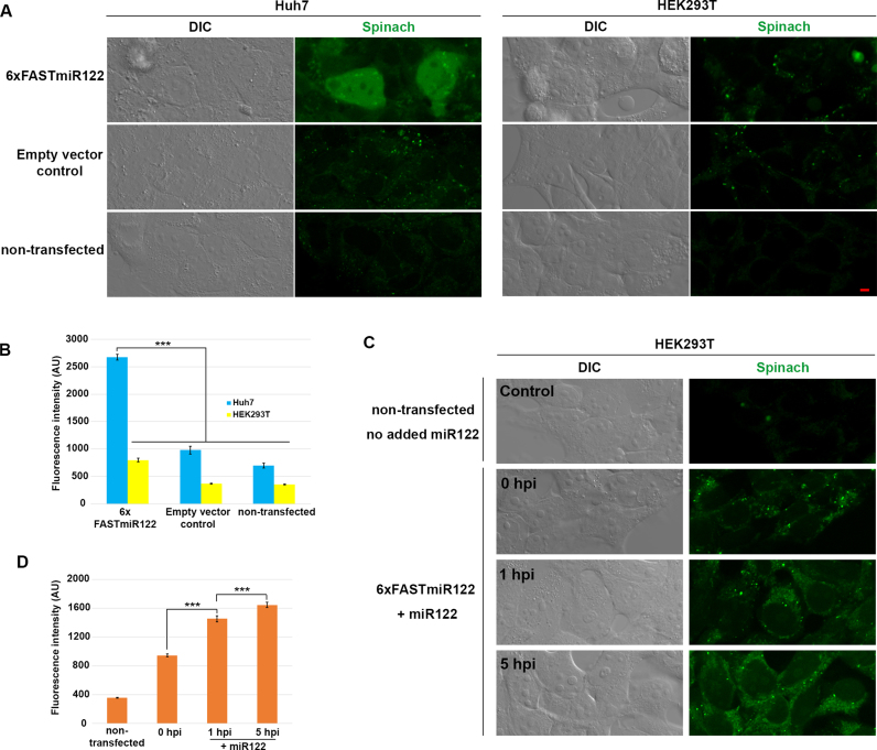
FASTmiR122 sensor localized miR122 and detects changes of miR122 in live mammalian cells. (A) The 6xFASTmiR122 sensor detected higher level of miR122 in nuclei and the cytosol of Huh7 cells (green, row 1). The empty vector (row 2) and non-transfected control (row 3) showed similar levels of background fluorescence. (B) Quantification of fluorescence in nuclei showed substantially stronger fluorescence intensity in Huh7 cells transfected with 6xFASTmiR122 sensors compared to the controls (n = 13). (C) HEK293T cells were transfected with the 6xFASTmiR122 sensor and miR122 was expressed using a lentivirus delivery system. Fluorescence increased 1 hpi (hours post infection) and 5 hpi compared with 0 hpi and cells without the 6xFASTmiR122 sensor (non-transfected control). (D) Quantification of nucleus-localized signal for miR122-expressing HEK293T cells (n = 15). Scale bars = 5 μM for all images. Error bars show standard error. Significance codes: ‘***’ for <0.001, ‘**’ for <0.01, ‘*’ for < 0.05, ‘NS’ for no significance (n = 10).

