Figure 5.
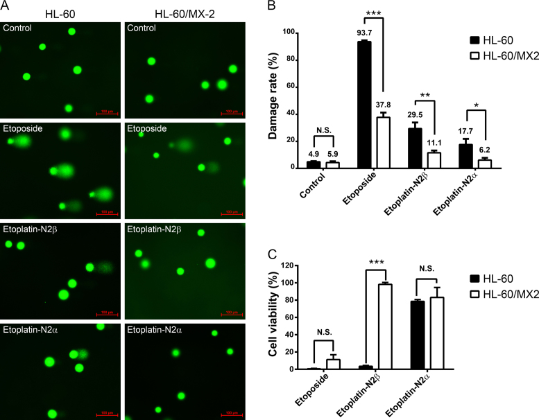
Like etoposide, etoplatins induced chromosomal DNA breaks and cancer cell death in a hTop2-dependent manner. (A) Levels of etoposide- and etoplatin-induced chromosomal DNA breaks are significantly reduced in the hTop2-deficient HL-60/MX2 cells compared to the parental HL-60 cells. It has been shown that HL-60/MX2, a mitoxantrone-resistant variant of HL-60 cell line, expresses much lower levels of both hTop2α and hTop2β (28). Cells were treated with 50 μM of etoposide or etoplatins for 1 h, comet assay were then carried out to measure chromosomal DNA breaks and quantified data were shown in (B). In the microscopic images, each green circular head or comet (head + tail) image represents one cell. The green dots are indicative of intact nucleoids containing SC loops of chromosomal DNA attached to the nuclear matrix. Upon drug treatments, damaged nuclear DNA becomes relaxed due to the presence of DNA breaks resulting from drug-stabilized Top2cc and a tail of damaged DNA migrates toward the anode during electrophoresis, leading to the comet-like appearance. Values as determined by the percentage of cells with DNA damage are presented as mean ± SD (n = 3). (C) Etoplatin-N2β exhibits clear hTop2-dependent cancer cell killing activity. HL-60 leukemia cells were treated with 100 μM of etoposide, etoplatin-N2β or etoplatin-N2α, and after 4 days of incubation, MTT assay were performed to determine cytotoxicity. N.S. (not significant, P > 0.05); *P < 0.05; **P < 0.01; ***P < 0.001.

