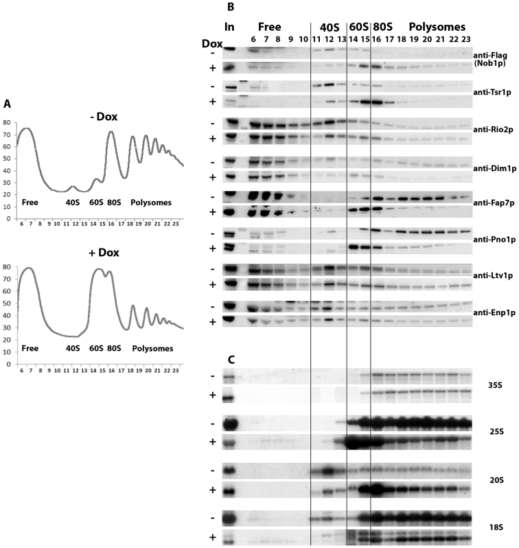
Figure 1.
Sucrose gradient fractionation of total extracts from a TetO7-RIO1, NOB1-FPZ strain grown in YPD or in YPD plus doxycycline for 16 h to repress RIO1 expression. (A) Recording of A254 nm profiles. Top: Rio1p-expressing cells (- Dox); bottom: Rio1p-depleted cells (+ Dox). The peaks of free proteins, free 40S and 60S subunits, 80S ribosomes and polysomes are indicated as well as fraction numbers. (B) Sedimentation profile of pre-40S particle AFs analyzed by western blot. Proteins were purified from the indicated fractions (top) of gradients loaded with extracts from Rio1p-expressing cells (–Dox) or Rio1p-depleted cells (+Dox). The antibodies used for the detection of specific factors are indicated. The anti-FLAG antibody allows the detection of tagged Nob1p. (C) Sedimentation profile of (pre)-rRNAs analyzed by northern. Same as in (B) except that RNAs were extracted from gradient fractions, separated by agarose gel electrophoresis in denaturing conditions, transferred to a nylon membrane and detected using various specific oligonucleotide probes.

