Figure 6.
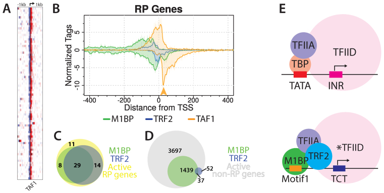
TAF1 is present at RP gene promoters. (A) Heatmaps display TAF1 ChIP-exo reads from S2R+ cells piled from −1 to +1 kb relative to RP gene TSS. Rows represent individual genes and are sorted by M1BP reads summed in a 2kb window as in Figure 5A. The TSS position is indicated by the arrow at the top of the panel. Duplicate genes were refined to a single isoform by removing eight paralogs lacking TRF2 or a TCT motif in the promoter. (B) Composite plots for M1BP, TRF2 and TAF1 were generated from the same RP gene list used for the heatmap. The orange arrow highlights a TAF1 peak that aligns with an M1BP peak. (C and D) Venn diagrams showing the overlap between M1BP and TRF2 peaks present at all active RP gene promoters (n = 62) or all other active gene promoters (n = 5225). (E) Model depicting M1BP’s recruitment of TRF2 at RP gene promoters. At TATA-containing promoters, TBP-bound TFIID engages with promoter sequences both up- and downstream of the TSS. At the majority of RP gene promoters, which lack both a TATA box and initiator sequence, M1BP and TRF2 bind the core promoter upstream of the TSS. The asterisk(*) denotes a noncanonical TFIID complex proposed to have TRF2 substituting for TBP.

