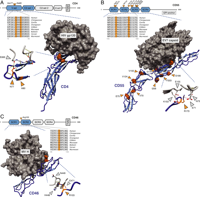
Figure 4.
Positively selected positions in the interaction between viruses and their cellular receptors. (A) CD4—HIV-1 envelop protein gp120 (2NXY (73)). (B) CD55 [DAF]—echovirus 7 capsid (3IYP (75)). (C) CD46 [MCP]—measles virus H (3INB (77)). (A, C) structures of interacting proteins crystallized together as complexes; (B) EM reconstruction fitted with the individual crystal structures. Hydrogen bonds are in yellow; charge interactions are shown as dashed lines (inset of B). Blue: human receptors, orange: PSR, gray: viral proteins. Protein domains that are present in the structures are in blue and marked by the dashed lines. PSR numbering is based on the human Ensembl sequence. Viral positions correspond to sequences represented in the structures.

