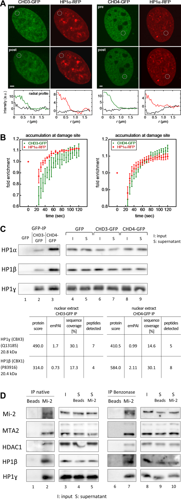
Figure 4.
CHD3 and CHD4 are rapidly recruited to laser-induced DNA repair sites and interact with HP1. (A) U2OS cells transiently expressing HP1α-RFP and CHD3/4-GFP were subjected to localized microirradiation with a 405 nm laser. CHD3-GFP, CHD4-GFP and HP1α-RFP accumulated at the damage site (gray circles). Radial intensity profiles around the damage site are shown below the images for clarity. Scale bar, 2 μm. (B) Accumulation kinetics of the indicated constructs at the damage site. (C) The expression of GFP (control), CHD3-GFP or CHD4-GFP in stably transfected Flp-In™ T-Rex™ 293 cells was induced by doxycycline treatment for 24 h. Subsequently, (i) whole cell extracts (WCE) and (ii) nuclear extracts (NE) were prepared and subjected to IP reactions, using GFP-Trap®_A beads. The IP reactions were finally analysed by (i) western blot with the indicated HP1 antibodies or (ii) mass spectrometry. The reactions were finally analyzed by mass spectrometry. A quantitative difference in protein amount levels is reflected by the emPAI-values (exponentially modified protein abundance index) (121). (*) Only one unique peptide was found in the mass spectrometry analysis (see also Materials and Methods). (D) Nuclear extract (NE) from non-transfected Flp-In™ T-Rex™ 293 cells was prepared and subjected to IP reactions in the absence or presence of Benzonase (see also Materials and Methods), using Protein A beads (+ Mi-2 antibody or no antibody as control). The IP reactions were finally analysed by western blot with the indicated HP1 and Mi-2-NuRD antibodies. Number of replicates: (A/B) (left) CHD3-GFP and HP1-alpha-RFP (12); (right) CHD4-GFP and HP1-alpha-RFP (12); (C) 3; (D) 3.

