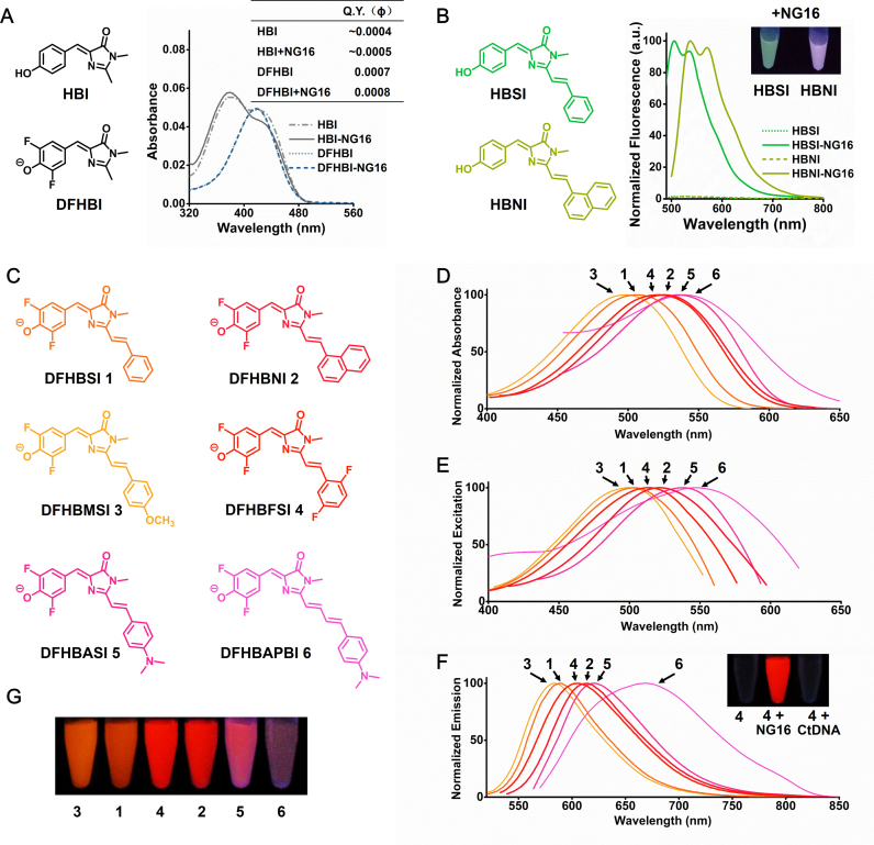
Figure 1.
(A) Absorption spectra and Q.Y. of HBI (3 μM) and DFHBI (3 μM) in the absence and presence of NG16 (30 μM). (B) NG16 (15 μM) enhances the fluorescence of HBSI (3 μM) and HBNI (3 μM). Inset is the photograph of HBSI and HBNI with NG16 under illumination of ultraviolet (UV, 365 nm) light. (C) Structures of the RFP chromophore analogues. (D) Absorption, (E) excitation and (F) emission spectra of chromophore–NG16 complexes (3 μM). Inset of F is the solutions containing DFHBFSI, DFHBFSI with NG16, and DFHBFSI with calf thymus DNA (CtDNA) photographed under illumination of UV light. (G) Chromophore-NG16 complexes were illuminated with UV light and photographed. The image is a montage obtained under identical image-acquisition conditions.

