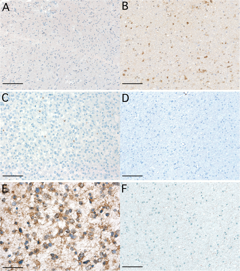
Fig. 1.
Difference in TIL density and PD-L1 expression in IDH-wt and IDH-mut glioma. (A, C, E) IDH-wt glioma WHO grade III without immunohistochemical staining for IDH-R132H mutant and lack of IDH gene 1 or 2 mutations based on gene sequencing (magnification × 100; A; scale bar 250 μm), scattered infiltration with CD3+ TILs (magnification × 200; C; scale bar 100 μm), fibrillary expression of PD-L1 (magnification × 400; E). (B, D, F) IDH-mut WHO grade II glioma presenting with anti–IDH-R132H immunostaining (magnification × 100; B), absence of CD3+ TILs (magnification × 200; D; scale bar 100 μm), lack of PD-L1 expression (magnification × 200; F; scale bar 100 μm).

