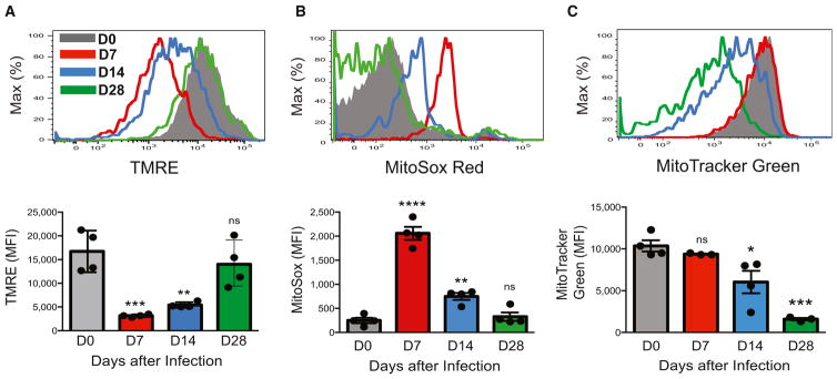
Figure 1. NK Cells Undergo Dynamic Changes in Mitochondrial Quality in Response to Viral Infection In Vivo.
WT splenic Ly49H+ NK cells were transferred into Ly49H-deficient mice and infected with MCMV. Shown are representative histograms and quantification of mean fluorescence intensity (MFI) for tetramethylrhodamine ethyl ester (TMRE) staining (A), MitoSox Red staining (B), or MitoTracker Green staining (C) in transferred NK cells from recipient spleens enriched with NK cells at the indicated time points after infection. Data are representative of three independent experiments with n = 3–4 per time point. Samples were compared with an unpaired, two-tailed Student’s t test, and data are presented as the mean ± SEM (*p < 0.05; **p < 0.01; ***p < 0.001; ****p < 0.0001).

