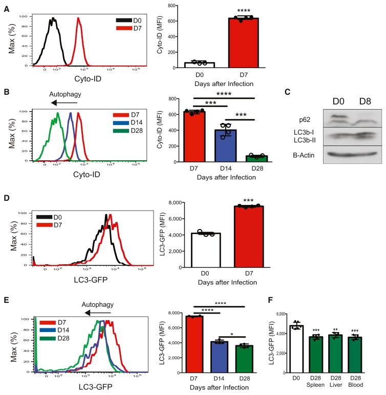
Figure 2. NK Cells Induce Autophagy in Response to Viral Infection In Vivo.
(A and B) WT splenic Ly49H+ NK cells were transferred into Ly49H-deficient mice and infected with MCMV. Shown are representative histograms and quantification of MFI for Cyto-ID staining of autophagosomes at each indicated time point after infection.
(C–F) LC3-GFP splenic Ly49H+ NK cells were transferred into Ly49H-deficient mice and infected with MCMV. (C) Representative Western blot of LC3b and p62 for adoptively transferred splenic Ly49H+ NK cells at day 8 after infection or for naive NK cells. (D–F) Representative histograms and quantification of LC3-GFP MFI of adoptively transferred Ly49H+ NK cells in the blood on days 0 and 7 after infection (D) and days 7, 14, and 28 after infection (E) and of indicated tissues of recipients on days 0 and 28 after infection (F).
Data are representative of three independent experiments with n = 3–7 per group per time point. Samples were compared with an unpaired, two-tailed Student’s t test, and data are presented as the mean ± SEM (*p < 0.05; **p < 0.01; ***p < 0.001; ****p < 0.0001). See also Figure S1.

