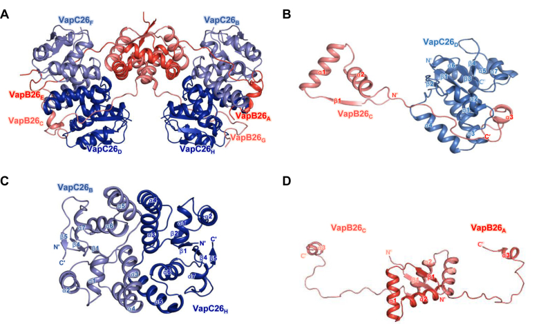
Figure 1.
Overall structures of VapBC26 and its subunits. (A) Ribbon representation of the VapBC26 hetero-octamer. Chains A and E are shown in red and chains C and G of VapB26 are shown in salmon. Chains B and F and chains D and H of VapC26 are shown in slate and blue, respectively. (B) Structure of the VapBC26 heterodimer. An arm of VapB26 covers VapC26. (C and D) Structure of (C) the VapC26 and (D) VapB26 dimer detached from the VapBC26 assembly. VapC26 displays cross-contacts between the α5 helix of one toxin monomer and the α3 and α4 helices of another proximal toxin monomer.

