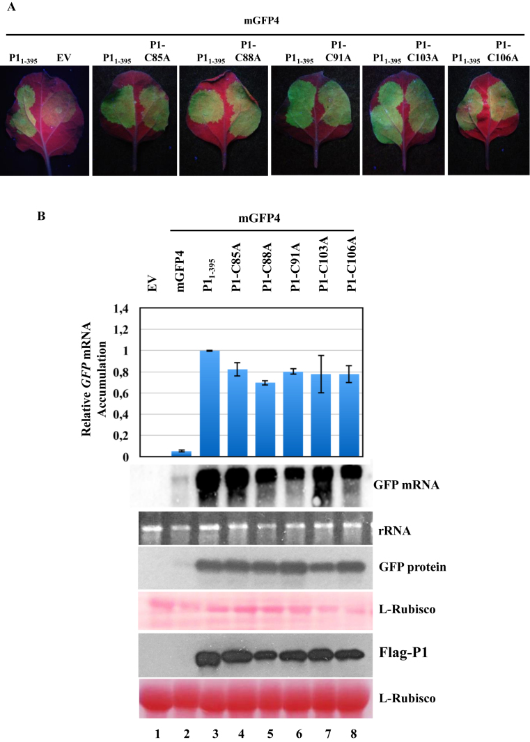
Figure 3.
Mutational analysis of the zinc finger domain present in SPMMV P1. (A) 35S:P1-C85A, 35S:P1-C88A, 35S:P1-C91A, 35S:P1-C103A and 35S:P1-C106A respectively were coinfiltrated with 35S:mGFP4. Photos were taken under UV light at 3 days post agroinfiltration. (B) GFP mRNA was detected by Northern blotting. Extracts of infiltrated leaves were subjected to SDS-PAGE followed by immunoblotting with the anti-GFP antibody. Flag-tagged P1 proteins were detected by Western blotting using the M2-anti Flag antibody. Ponceau staining shows equal loading of proteins in both Western blots. Top panel, mean (n = 3) relative mGFP4 transcript level and ±SD (lane 3 = 1.0 for mGFP4 transcript). One representative blot from three biological replicates is shown.

