
Figure 3.
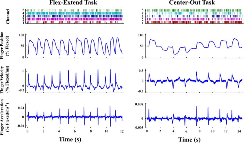
Finger kinematics and associated neural spikes from (left) Monkey P performing the flex-extend task and (right) Monkey L performing the center-out task. Each spike raster displays five separate channels, chosen to be exemplary of modulated activity in each monkey. Within a raster row, the time of each individual spike event is represented by a tick.