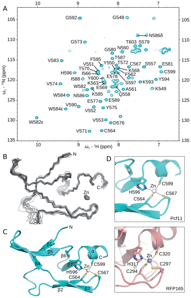
Figure 4.
Solution structure of the second zinc-binding domain. (A) 1H,15N HSQC of [15N]-labelled Pcf11 (530-626) in 50 mM Tris (pH 7.5) and 150 mM NaCl, collected at 298 K. Crosspeaks corresponding to residues G548 to T603 have been annotated. (B) Backbone traces for the ensemble of 15 structures calculated for residues 548–603. The Zn2+ ion is displayed as a sphere. (C) Cartoon representation of the structure closest to the mean, with secondary structure elements labelled and the Zn2+ ion shown as a sphere. The zinc-binding residues are shown in stick form and are labelled. (D) Comparison of the local region of structure similarity between the second zinc-binding domain of Pcf11 (top) to only one of the two zinc-binding sites of the RING finger proteins (bottom), in this case from RFP165 (PDB 5D0I; (50)).

