Figure 3.
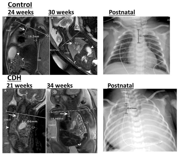
Coronal T2-weighted MRI of a control fetus and fetus with congenital diaphragmatic hernia.
Top left: A: 24 weeks and B: 30 week images in the same fetus demonstrating the normal appearance of the chest. The horizontal line at the level of the thoracic inlet (TI) and vertical line represents the distance from the TI to the carina (white arrow). Normal lungs are seen (open arrow) as well as normal position of the stomach (white arrowhead) and liver (curved arrow).
Bottom left: A: 21 week and B: 34 week images showing left sided congenital diaphragmatic hernia. hernia (open arrow). Horizontal line shows the level of the thoracic inlet and short vertical line measures the distance from the thoracic inlet to the carina pointed by a large white arrow. The herniated viscera (open arrow) and right lung (arrow head) are seen. B: Same fetus at 34 weeks gestational age shows similar calculation of the distance between the thoracic inlet and the carina (white arrow). Bowel loops are seen herniated into the left chest (open arrow) and the liver is seen in the abdomen (curved arrow).
Top right: Anterior-posterior radiograph of the chest in control infant demonstrating the normal appearance of the chest. The vertical line at the level of clavicles represents the distance from the thoracic inlet to the carina.
Bottom right: Anterior-posterior radiograph of the chest in an infant with left sided congenital diaphragmatic hernia demonstrating herniation of the abdominal viscera into the chest. The vertical line at the level of clavicles represents the distance from the thoracic inlet to the carina.

