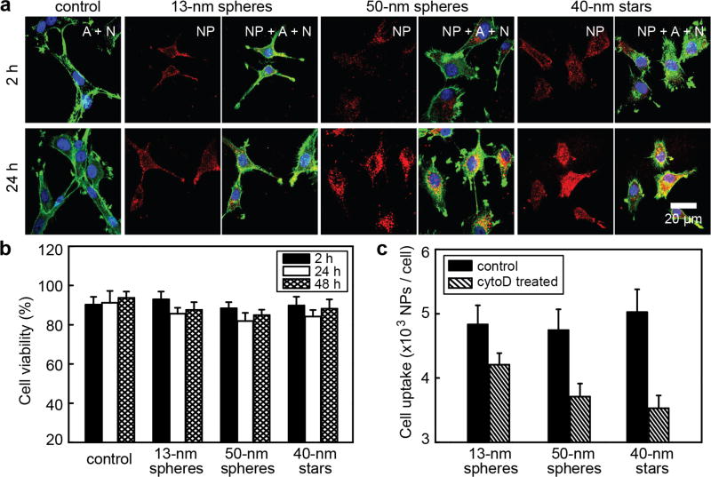
Figure 5.
Intracellular distribution of NP-siRNA constructs and cell morphology changes induced by larger constructs. (a) Confocal fluorescence images of cells treated with PBS (control) or three different formulations of constructs with the same concentration of NPs (0.2 nM); The actin (A) cytoskeleton and nucleus (N) were stained with Alexa Fluor® 594 Phalloidin (green) and DAPI (blue), respectively; nanoconstructs were labeled with Cy5 (red). The scale bar is 20 µm for all images. (b) MTS assay of the cell viabilities of U87 cells treated with three different formulations of NP-siRNA/Cy5 (0.2 nM) or PBS as the control; (c) Cell uptake inhibition by CytoD for three formulations. Following 30-min pre-treatment with CytoD, U87 cells were incubated with NP-siRNA in presence of CytoD for another 30 min and cell uptake was measured by ICP-MS. As a control, U87 cells were also treated with constructs under the same conditions, but without cytoD.

