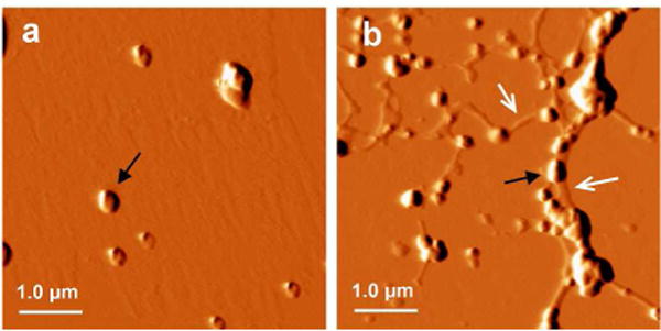
Figure 2.

Tapping mode AFM images of AuNPs in the absence or presence of preformed Aβ40 amyloids. The AuNPs were incubated in pH 7.4 phosphate buffer alone (a), or with additional 1.7 μM Aβ40 amyloids (b). The black arrow indicates the AuNP cluster. The white arrow indicates the fibrils.
