Figure 6.

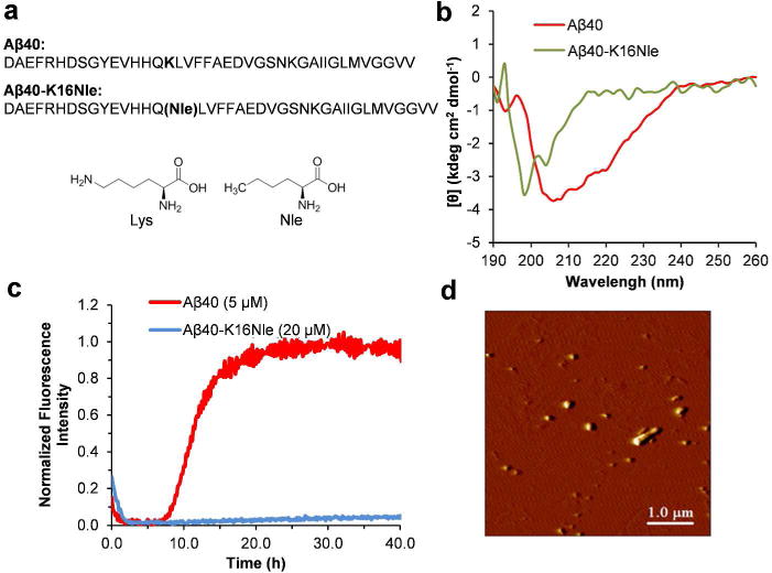
Aggregation of Aβ40-K16Nle. (a) The primary sequence of Aβ40 and Aβ40-K16Nle. The chemical structures of Lys and Nle are shown for comparison. (b) CD spectra of Aβ40 (50 μM) and M1 (40 μM) after 5 d incubation in pH 7.4 phosphate buffer at 37°C. (c) Aggregation kinetics of Aβ40 and Aβ40-K16Nle followed by ThT fluorescence in pH 7.4 phosphate buffer at 37°C. (d) AFM image of Aβ40-K16Nle (40 μM) incubated for 5 d in pH 7.4 phosphate buffers at 37°C.
