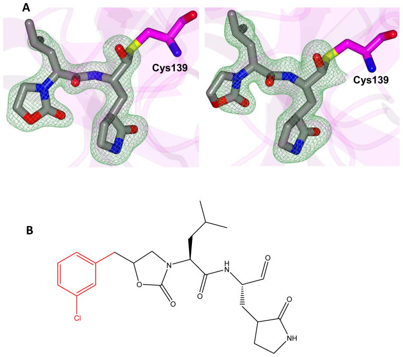
Figure 2.
A) Fo-Fc polder omit map [53] of inhibitor 8B (green mesh) contoured at 3σ. The ligand associated with subunits A and B is positioned on the left and right, respectively. B) Structure of inhibitor 8B with the disordered m-chlorobenzyl group highlighted in red.

