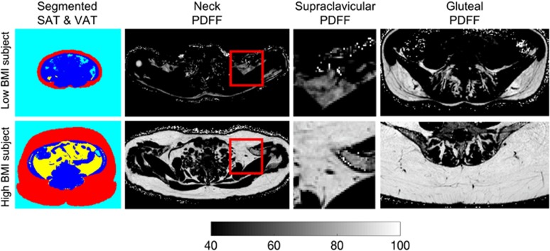
Figure 1.
Comparison of different fat depots in a low BMI female subject (first row, BMI: 17.4 kg m–2, age=39 years) and a high BMI female subject (second row, BMI: 38.1 kg m–2, age=48 years). The high BMI subject has higher SAT volume and higher VAT volume (color-coded masking using red for SAT, yellow for VAT, blue for non-adipose tissue and cyan for air on first column), higher supraclavicular PDFF (full-axial and zoomed PDFF maps on second and third columns) and higher gluteal PDFF (gluteal PDFF maps on fourth column) than the low BMI subject.

