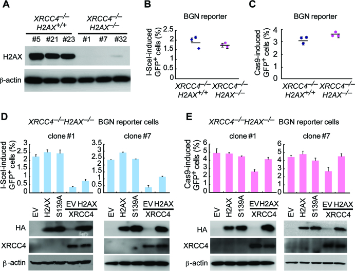
Figure 5.
XRCC4 mediates H2AX-dependent NHEJ. (A) Deletion of H2AX in XRCC4–/– BGN reporter cells by CRISPR/Cas9. XRCC4–/– BGN reporter mouse ES cells were transfected twice with Cas9 expression plasmids and H2AX gRNA expression plasmids and then plated on MEF. In about 2 weeks, individual clones were picked, and deletion of H2AX was identified by Western blot (upper). (B and C) Percentage of I-SceI (B) and Cas9 (C)-induced GFP+ cells from XRCC4–/–H2AX+/+ and XRCC4–/–H2AX–/– BGN reporter cells. Bars represent the mean ± S.D. of three independent experiments, each performed in triplicates. One-way Anova between ‘XRCC4–/–H2AX+/+’ and ‘XRCC4–/–H2AX–/–’: NS. (D and E) Percentage of I-SceI (D) and Cas9 (E)-induced GFP+ cells from two XRCC4–/–H2AX–/– BGN reporter cell clones transiently transfected with mouse H2AX and/or XRCC4 expression plasmids. Bras represent the mean ± S.D. of three independent experiments, each in triplicates. In I-SceI-induced NHEJ assays (D), paired t-test in clone#1 (left): NS between ‘EV’ and ‘H2AX’; P = 0.005 between ‘EV’ and ‘EV+XRCC4’; and P = 0.024 between ‘EV+XRCC4’ and ‘H2AX+XRCC4’. Paired t-test in clone#7 (right): NS between ‘EV’ and ‘H2AX’; P = 0.001 between ‘EV’ and ‘EV+XRCC4’; and P = 0.007 ‘EV+XRCC4’ and ‘H2AX+XRCC4’. In Cas9-induced test (E), paired t-test in clone#1 (left): NS between ‘EV’ and ‘H2AX’; P = 0.047 between ‘EV’ and ‘EV+XRCC4’; and P = 0.002 between ‘EV+XRCC4’ and ‘H2AX+XRCC4’. Paired t-test in clone#7 (right): NS between ‘EV’ and ‘H2AX’; P = 0.034 between ‘EV’ and ‘EV+XRCC4’; and P = 0.035 between ‘EV+XRCC4’ and ‘H2AX+XRCC4’. Expression of exogenous H2AX (HA-tagged) and XRCC4 was detected by western blot as indicated.

