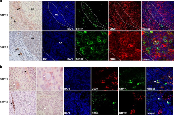
Figure 2.
Expression of S1PR1 and S1PR2 in normal lymphoid tissues and HRS cells. (a) Normal tonsil. Upper panels: S1PR1 expression in MZ B cells, but not in GC B cells, confirmed by costaining for CD20. Lower panels: S1PR2 was expressed in normal GC B cells, confirmed by costaining with CD20 (white arrow) and BCL6 (Supplementary Figure S4). (b) HRS cells expressed S1PR1 but not S1PR2 (white arrows) confirmed by CD30 costaining. Endothelial cells and red blood cells were strongly positive for S1PR1 and S1PR2, respectively (black arrows). Further examples of S1PR1 staining in HRS cells indicating the presence of both membrane and cytoplasmic staining are shown in Supplementary Figure S5.

