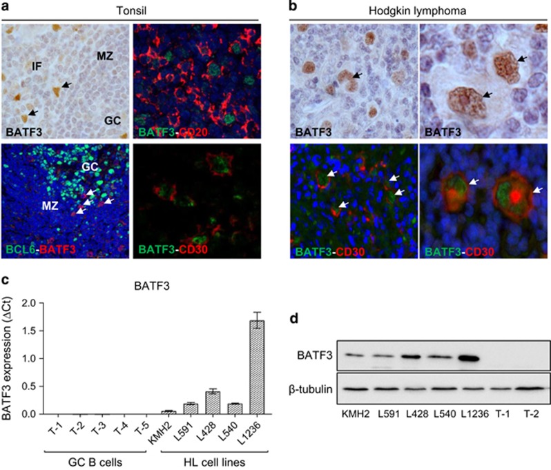
Figure 5.
BATF3 is overexpressed in HRS cells. (a) IHC of normal tonsil. BATF3-positive cells were mainly located outside the GC, of which some expressed CD20 and CD30 but not BCL6 (arrowed). (b) BATF3 expression in CD30-positive HRS cells (arrowed). (c) BATF3 mRNA and (d) protein expression in HL cell lines. T1-T5 are GC B cells isolated from five donors. IF, interfollicular; MZ, mantle zone.

