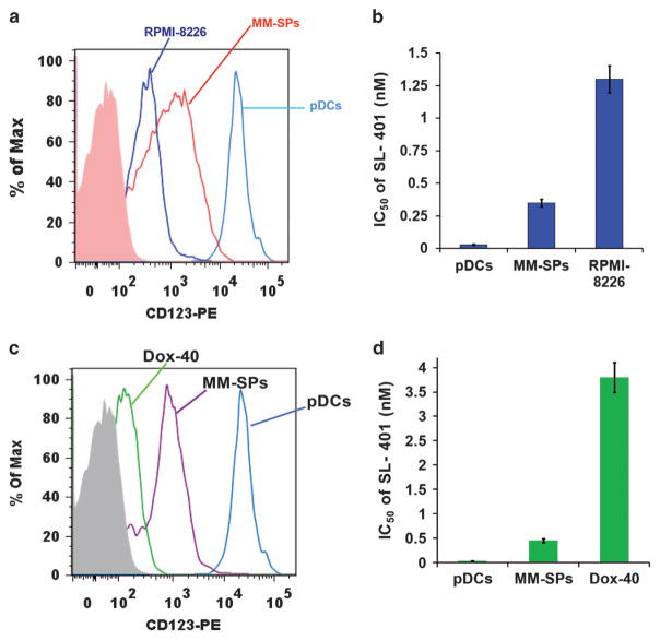
Figure 5.
Comparative analysis of SL-401 activity against clonogenic MM side population (MM-SPs) and MM cells (a) MM-SPs were isolated from RPMI-8226 using Hoechst 33342 staining (described in methods; ~ 99% purity). pDCs, MM-SPs and RPMI-8226 cells were analyzed for IL-3 R expression using anti-IL-3Rα-PE Abs and flow cytometry (mean ±s.d.; n =5). (b) RPMI-8226, MM-SPs and pDCs were treated with increasing concentrations of SL-401 for 48 h, and then analyzed for viability by WST assay. Bar graph shows IC50 of SL-401 against RPMI-8226, MM-SPs and pDCs (mean ±s.d.; n =5; P<0.005). (c) MM-SPs were isolated from RPMI-8226 R/Dox-40 using Hoechst 33342 staining (described in methods; ~ 99% purity). pDCs, MM-SPs and Dox-40 cells were analyzed for IL-3 R expression using anti-IL-3Rα-PE Abs and flow cytometry (mean ±s.d.; n =5). (d) Dox-40, MM-SPs, and pDCs were treated with increasing concentrations of SL-401 for 48 h and then analyzed for viability by WST assay. Bar graph shows IC50 of SL-401 against RPMI-8226 R/Dox-40, MM-SPs and pDCs (mean ± s.d.; n = 5; P <0.005).

