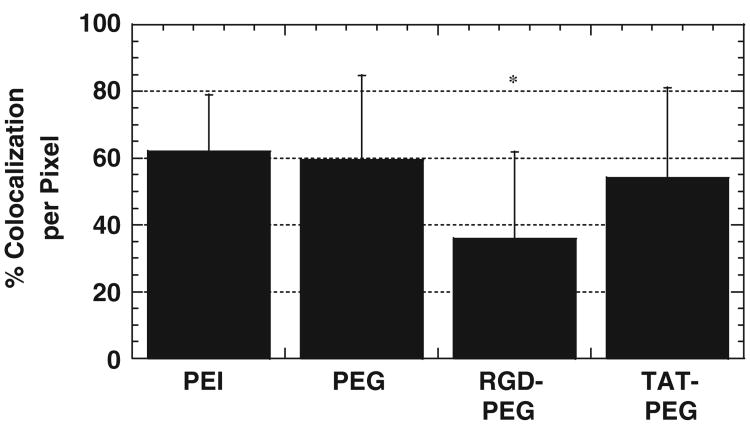
Fig. 5.
Percentage of co-localization between PEI/DNA nanocomplexes (N/P = 20) and acidic vesicles at 12 h post-transfection, measured by numbers of overlapping pixels (n = 50 cells). PEI–PEG–RGD/DNA (RGD–PEG) displays lowest co-localization, and the difference is statistically significant (*) (p<0.01, ANOVA) compared to all other complexes.

