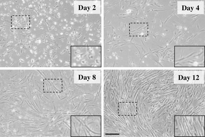
Fig 1. Representative images of gilthead sea bream cultured myocytes at days 2, 4, 8 and 12 of development.
Images were taken with an EOS 1000D Canon digital camera coupled to an Axiovert 40C inverted microscope (Carl Zeiss, Germany). Objective: 10x. Scale bar: 50 μm. Insets in each image are enlarged views of cells from each panel.

