Figure 1.

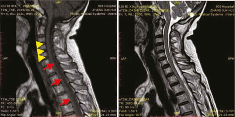
Magnetic resonance imaging shows multiple disc herniation of cervical 3/4, 4/5, and 5/6. The epidural sac is compressed by disc herniation (yellow arrow head). Epidural inflammation is seen with increased signal intensity (red arrow).

Magnetic resonance imaging shows multiple disc herniation of cervical 3/4, 4/5, and 5/6. The epidural sac is compressed by disc herniation (yellow arrow head). Epidural inflammation is seen with increased signal intensity (red arrow).