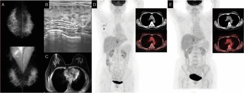
Figure 4.

Mammography (A), breast ultrasound (B), and magnetic resonance imaging (C) do not show abnormalities in the breast. Positron emission tomography (D) shows abnormal increased FDG uptake in right axillary region. Positron emission tomography (E) does not show abnormal increased FDG uptake in the right axillary region after neoadjuvant chemotherapy. FDG= flurodeoxyglucose.
