
Fig. 5.
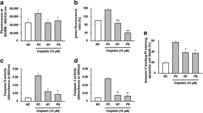
Antioxidant effect of NY and PS on cisplatin-induced cell death in HK-2 cells. Cells were treated with 10 μM cisplatin for 30 m and the level of ROS was detected by microplate reader (a). Cells were exposed to cisplatin for 24 h with or without PS or NY (b). The measures of JC-1 fluorescence intensity (c and d). Cells were treated to bind Annexin V and then to undergo PI staining; exposed to cisplatin for 24 h with or without PS or NY. HK-2 cells were treated with cisplatin for 24 h to measure apoptosis (e). Cell lysates were assayed for caspase-9, 3 activity. The statistical significance (*p < 0.05, **p < 0.01 and ***p < 0.001) of the observed differences between the cisplatin-treated and herbal groups was determined by a t-test