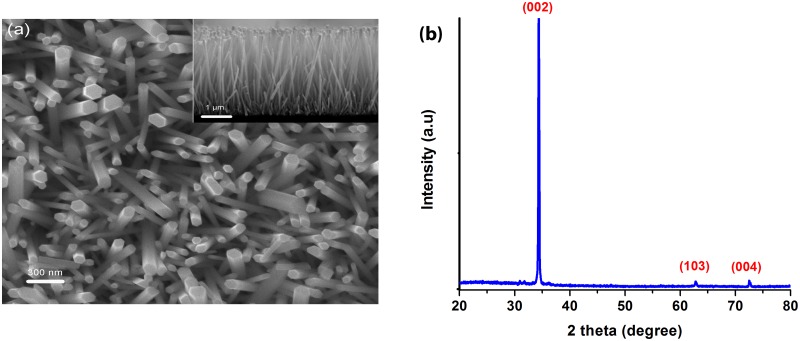
Fig 2. (a) Top and cross-sectional (inset) SEM micrographs and (b) XRD pattern of ZnO nanorods.
(a) SEM micrographs of ZnO nanorods and (b) XRD pattern of the microwave assisted hydrothermally grown ZnO nanorods on glass substrate. Samples were annealed at 350°C for 1 h after the hydrothermal growth.

