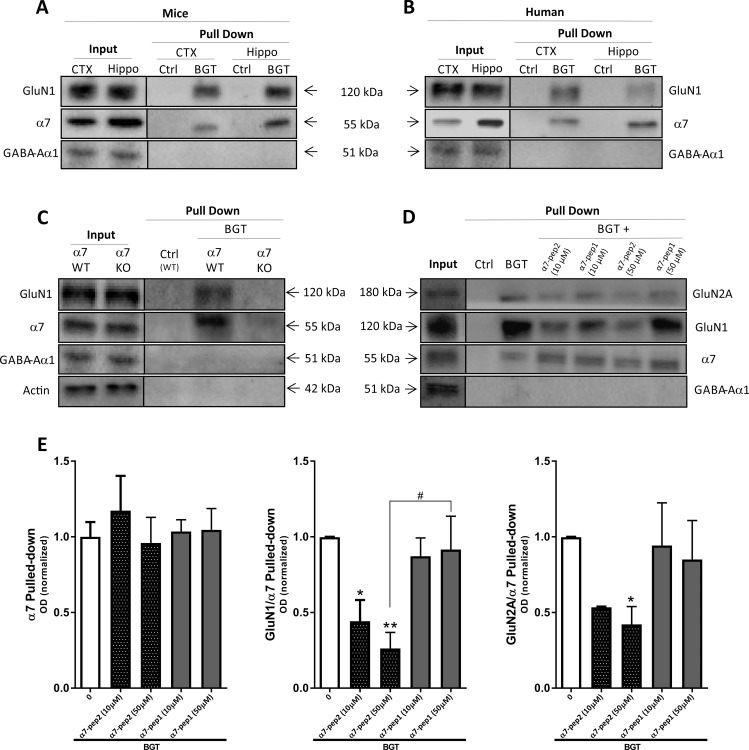
Fig 1. Complex formation between α7 nAChR and NMDAR in murine and human cortex and hippocampus.
(A-B) Affinity purification with agarose beads covalently coupled with α-bungarotoxin (BGT) or BSA (Ctrl) on homogenates from murine (A) and human (B) cortical (CTX) and hippocampal (Hippo) tissues. Total lysates (Input) and pulled-down (Pull Down) samples were submitted to gel electrophoresis and Western blotting followed by detection using antibodies for GluN1, α7 nAChR and GABAAR α1 subunits. The gels in A and B are representative for different experiments using tissues from 4 different mouse hippocampi, 4 different mouse cortices, 2 different human hippocampi and 2 different human cortices. (C) Total lysates (Input) and pulled-down (Pull Down) samples from WT and α7 KO mouse cortical homogenates were submitted to gel electrophoresis and Western blotting followed by detection using antibodies for GluN1, α7 nAChR and GABAAR α1 subunits and β-actin. (D) Total lysates (Input) and pulled-down (Pull Down) samples from mouse cortical homogenates pretreated with buffer or buffer supplemented with α7-pep2 (10 μM and 50 μM) or α7-pep1 (10 μM and 50 μM) were submitted to gel electrophoresis and Western blotting followed by detection using antibodies for GluN1, GluN2A, α7 nAChR and GABAAR α1 subunits. (E) Quantification of α7 pulled-down and GluN1 and GluN2A pulled-down (normalized to the pulled-down α7) from mouse cortical homogenates pretreated with buffer or buffer supplemented with α7-pep2 (10 μM and 50 μM) or α7-pep1 (10 μM and 50 μM). Values are given as mean ± SEM (n = 3–4, i.e. 3–4 different mouse cortices, the experiment was performed once). *p <0.05 and **p < 0.01 indicate statistically significant difference from the vehicle-treated group in Kruskal-Wallis test with Dunn’s multiple comparison test. #p <0.05 indicates statistically significant difference between GluN1/α7 Pulled-down ratios between α7-pep2 (50 μM) or α7-pep1 (50 μM) in unpaired t-tests.

