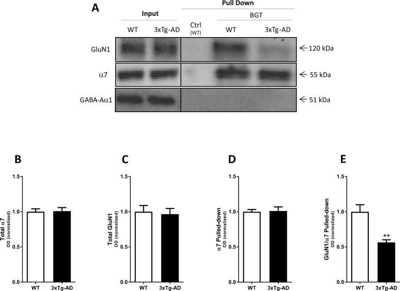
Fig 3. Complex formation between α7 nAChR and NMDAR in the adult 3xTg-AD mouse brain.
Affinity purification performed with agarose beads covalently coupled with α-bungarotoxin (BGT) or BSA (Ctrl) using frontal cortical tissue lysates from adult 3xTg-AD mice (76–84 weeks old) and age- and sex-matched WT mice. (A) A representative example of a western blot illustrating GluN1, α7 nAChR and GABAAR α1 protein levels in total lysates (Input) and pulled down (Pull Down) samples from WT and 3xTg-AD mouse cortical homogenates. (B-C) Quantification of total GluN1 (B) and total α7 (C) in lysates from WT and 3xTg-AD mouse cortical homogenates (both normalized to stain-free gel). (D-E) Quantification of α7 pulled-down (normalized to stain-free gel) (D), and of GluN1 pulled-down with α7 (normalized to the pulled-down α7) (E). In B-E, the control group (WT) is set to 1, and values are shown as mean ± SEM. **p < 0.01 indicates statistical significant difference from WT group in unpaired t-tests, n = 8 (WT) and n = 8 (3xTg-AD).

