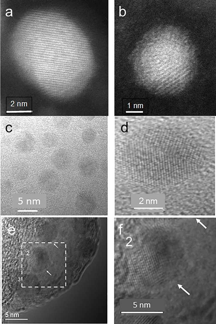
Fig 2. Electron microscopy images of Zn, ZnPEG400, and ZnPEG1000 nanoparticles.
TEM images (a, c, d) are showing the primarily-elemental zinc nanoparticles at different magnifications, (b) ZnPEG400 nanoparticle showing the metal core and the PEG passivation layer, (e) ZnPEG1000 nanoparticles with negative staining, and (f) ZnPEG1000 magnified from (e). The numbers 1, 2, 3 point to the nanoparticles with visible lattice fringe patterns, indicating their crystallinity.

