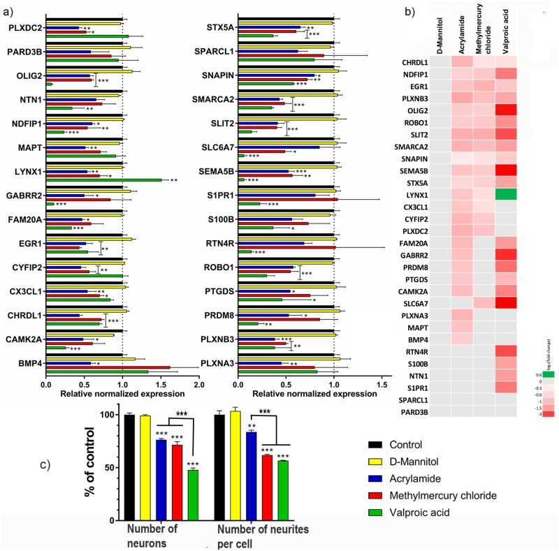
Fig 7. The effect of D-mannitol (negative control), acrylamide (ACR), methylmercury chloride (MeHg) and valproic acid sodium salt (VPA) on gene expression, the number of neurons and neurites per cell in differentiating C17.2 cells.
a) RT-qPCR of all 30 genes after 10 days of differentiation and exposure to the IC10 of said compounds (70 μM of ACR, 90 nM of MeHg and 100 μM of VPA. D-mannitol did not show any cytotoxicity for the concentrations used, and 1 mM was chosen for cellular exposure) b) Heatmap of the 30 genes expression during exposure to the 4 compounds. The log2(fold change) for the contrasts as compared to the control (unexposed) are illustrated c) the number of neurons and the number of neurites per cell decreased after exposure to all 3 neurotoxic compounds. The data are presented as the mean of 3 independent experiments performed in duplicates. Results were analyzed using two-way ANOVA followed by Dunnett’s multiple comparisons test. The bars represent the mean ± SEM. *p ≤ 0.05, **p ≤ 0.01, ***p ≤ 0.001 compared to control (cells exposed to only cell medium) or between the 3 different compounds (ACR, MeHg and VPA).

