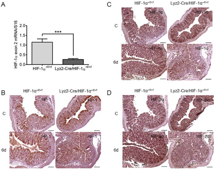
Fig 1. Hypoxia and HIF-1 accumulation after DSS-treatment.

Real time PCR of HIF-1α exon 2 (A) in RNA samples of hypoxic treated BMDM of wild type (HIF-1α+f/+f) and knockout (Lyz2-Cre/HIF-1α+f/+f) mice with 14 samples/group. Each bar represents the mean value ± SEM. ***P < 0.001. Immunohistochemical staining of hypoxia with Hypoxyprobe-1 (HP-1) antibody (B) or staining of HIF-1α (C) and HIF-2α (D) in paraffin-embedded colon tissue of wild type (HIF-1α+f/+f) and knockout (Lyz2-Cre/HIF-1α+f/+f) mice after treatment with drinking water (C = control) or with 2.5% DSS for six days (6d). Note, that the HIF-1α antibody recognizes both wild type and knockout HIF-1α (lacking exon 2) and therefore detects (non-functional) HIF-1α protein in knockout mouse tissue. Representative images of stained colon slices of experiments with five or six mice/group. Original bars 100 μm.
