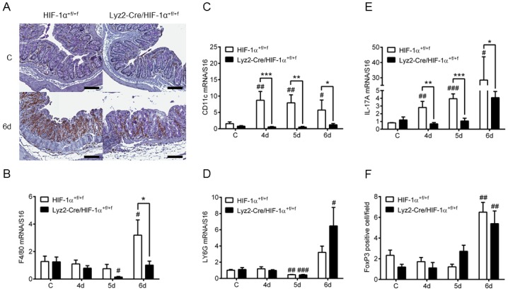
Fig 4. Accumulation of macrophages in colon tissue of mice lacking myeloid HIF-1α after DSS-treatment.

(A) F4/80 staining of paraffin-embedded colon sections from wild type (HIF-1α+f/+f) and knockout (Lyz2-Cre/HIF-1α+f/+f) mice treated with drinking water (C) or with 2.5% DSS (6d) for six days (6d). Real time PCR of markers for macrophages (F4/80) (B), dendritic cells (CD11c) (C), neutrophils (LY6G) (D) and Th17 cells (IL-17A) (E) in colon RNA samples of wild type (HIF-1α+f/+f) and knockout (Lyz2-Cre/HIF-1α+f/+f) mice treated for four to six days (4d-6d) with 2.5% DSS. (F) Number of FoxP3 positive cells/field of view in 4 fields/colon section of wild type (HIF-1α+f/+f) and knockout (Lyz2-Cre/HIF-1α+f/+f) mice treated as in (B). Data are representative for experiments with five or six mice/group. Each time point represents the mean value ± SEM. *P < 0.05; **P < 0.01 and ***P < 0.001 compared as indicated. # displays significance to respective control. Original bars, 100 μm.
