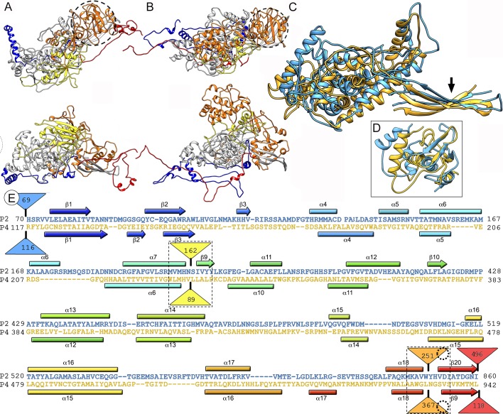
Fig 4. Structural similarities of P2 and P4 capsid proteins.
(A, B) Atomic models of P2 (A, top view, top; side view, bottom) and P4 (B, top view, top; side view, bottom). Conserved domains are grey (SID of P2 and P4, or P2* and P4*); insertions at the N terminus (blue), middle (yellow and orange), and C terminus (red) are also shown (the last 54 residues of P2 are omitted). Dashed circles correspond to P2 and P4 SIID. (C) Superimposed P2* (blue) and P4* (yellow) (non-superimposable regions for both CP are omitted). Arrow indicates shared β-sheet. (D) The P2 Cys738-Ser848 and the P4 Gly817-Thr929 segments (SIID of P2 and P4), included in the middle insertion (orange), are structurally aligned. (E) Sequence alignment of P2* (blue) and P4* (yellow) resulting from Dali structural alignment. α-helices (rectangles) and β-strands (arrows) are rainbow-colored from blue (N terminus) to red (C terminus) for each protein. Dashed rectangles indicate favorable insertion sites, triangles represent non-aligned segments (sizes indicated), and dashed circles correspond to P2 and P4 SIID. Insertions in sites 1 and 2 are shown as yellow and orange triangles, respectively.

