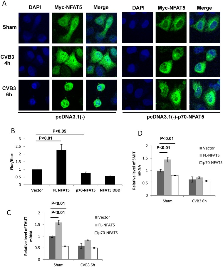
Fig 5. p70-NFAT5 acts as a dominant negative fragment of NFAT5.
(A) HeLa cells were co-transfected with pEGFP-myc-NFAT5 and pcDNA3.1(-)-p70-NFAT5 or pcDNA3.1(-) empty vector. Then the cells were infected with CVB3. At 4 and 6 h pi, the cells were fixed and immunostained using a specific antibody against the myc tag and observed by confocal microscopy. (B) HeLa cells were co-transfected with the luciferase reporter constructs pGL-TonE-luciferase or pRL-polIII and the plasmids expressing FL NFAT5, p70-NFAT5, NFAT5 DBD or pEGFP empty vector. At 48 h pt, the cell lysates were subjected to dual luciferase assay detecting the activity of the firefly luciferase (Fluc) and the renilla luciferase (Rluc). The relative activity (FLu/RLu) was determined after normalization against the vector control. (C, D) HeLa cells were transfected with plasmids expressing FL NFAT5 or p70-NFAT5 and then infected with CVB3. At 6 h pi, the cellular RNAs were extracted for qPCR measurement of mRNA level of TauT (C) and SMIT (D) (normalized to GAPDH as described in Fig 1). Vector-transfected cells were used as a control. Three biological replicates were performed for each assay and the results was subjected to statistical analysis.

