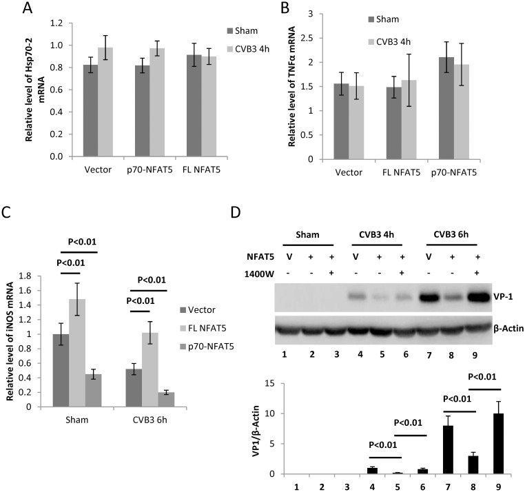
Fig 6. iNOS is essential for the anti-CVB3 effect of NFAT5.
HeLa cells were transfected with plasmids expressing FL NFAT5 or p70-NFAT5 and then infected with CVB3. At 6 h pi, the cellular RNAs were extracted for qPCR measurement of mRNA levels of Hsp70-2 (A), TNFα (B) and iNOS (C) (normalized to GAPDH as described in Fig 1). (D) HeLa cells transfected with pEGFP empty vector (V) or pEGFP-myc-NFAT5 were treated with 4 mM 1400W or DMSO control and then infected with CVB3 for VP1 detection and quantification (lower panel) as described in Fig 4A. Three biological replicates were performed for each assay and the result was subjected to statistical analysis.

