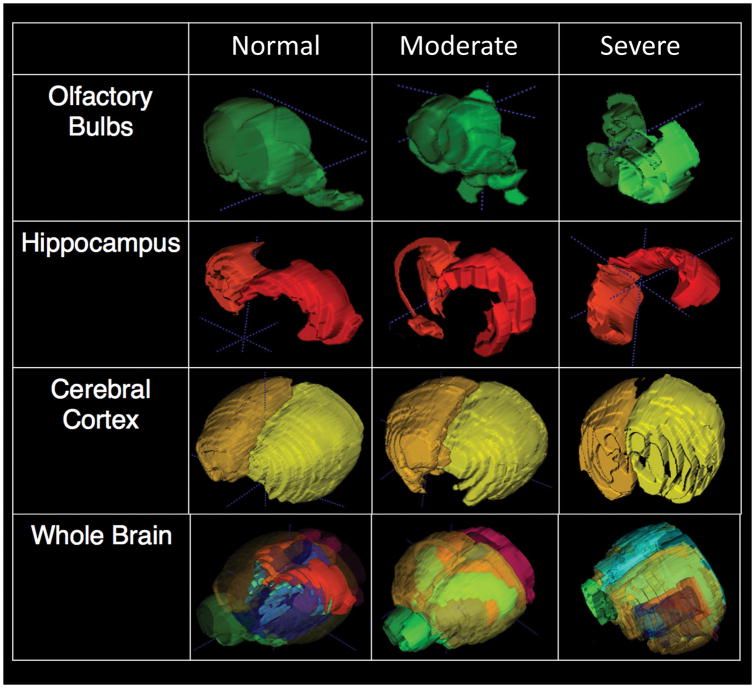
Figure 4.
Volumetric and morphometric analysis of the brain from the in vivo T2-weighted MRI. (left) normal WT mouse; (middle) homozygous mutant mouse with moderate hydrocephalus; (right) homozygous mutant mouse with very severe hydrocephalus. Examples of segmentation of different regions of interest (ROS) are shown for olfactory bulb, hippocampus, cerebral cortex, and the combined ROIs of the whole brain.

