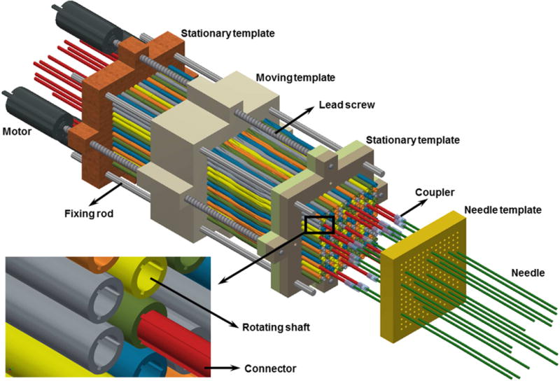
Figure 2.

Angular drive mechanism incorporating side views and cross sections of several points along the axis of a single needle. Translational motion of the moving template rotates the shaft, connector, and source/shield/catheter from (a) 0° to (b) 180° angular positions. Each needle, implanted through the patient template, is coupled to the catheter-mounted afterloader wire through a keyed connector (red), which passes through a rotating shaft. The catheter is rigidly attached to a proximal keyed cuff that enables the angular orientation of the shielded source to be fixed and known at all times during treatment. Items in the figure are not to scale.
